Campbell de Morgan spot
Granskad av Dr Toni Hazell, MRCGPSenast uppdaterad av Dr Rachel Hudson, MRCGPLast updated 31 Oct 2024
Uppfyller patientens redaktionella riktlinjer
- Ladda nerLadda ner
- Dela
- Language
- Diskussion
- Ljudversion
- Add to preferred sources on Google
Medicinska yrkesverksamma
Professional Reference articles are designed for health professionals to use. They are written by UK doctors and based on research evidence, UK and European Guidelines. You may find one of our hälsoartiklar more useful.
I den här artikeln:
Synonyms: cherry haemangiomas, senile angiomas
Fortsätt läsa nedan
What are Campbell de Morgan spots?
Campbell de Morgan spots, also known as cherry angiomas, are common, benign skin lesions of middle to older age, formed by proliferating, dilated capillaries and postcapillary venules. They are named after an English surgeon, Campbell de Morgan (1811-76).
Causes of Campbell de Morgan spots (aetiology) 1 2
Tillbaka till innehållTheir cause remains unknown:
Single studies have reported increased incidence in tropical climates, diabetes, transplant patients and those who are immunocompromised.
Pregnancy and prolactinomas are associated with the development of lesions, implicating hormonal mediators.
Numbers increase with age, so factors associated with the ageing process may be relevant.
Chemical exposure (mustard gas, 2-butoxyethanol) causes multiple lesions to develop.
Fortsätt läsa nedan
How common are Campbell de Morgan spots? (Epidemiology)1 2
Tillbaka till innehållThese are the most common cutaneous vascular proliferation. Few reports have been published recently but it is thought as many as 75% of those over 75 years old may have them.
They increase in frequency and size with age.
They increase in frequency from the age of 40.
They may occur anywhere but are most commonly found on the trunk.
They are seen across all races and sexes.
Visual appearance
Tillbaka till innehållCherry angioma on adult's arm
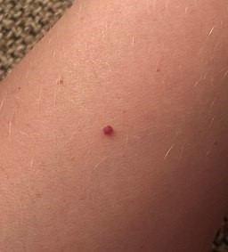
© Midasblenny, CC BY-SA 4.0, via Wikimedia Commons
1-3 mm diameter macules which may become larger papules over time.
Typical bright cherry red colour but can appear blue or purple.
They are non-blanching.
Fortsätt läsa nedan
Presentation
Tillbaka till innehållThey usually occur on the trunk and upper extremities.
They can be found at any skin site except the mucous membranes. The scalp has been reported.1
Lesions may be widespread, especially in the elderly.
They are usually asymptomatic.
Differentialdiagnos
Tillbaka till innehållThe diagnosis is usually clear clinically. Differential diagnosis may include:
Angiokeratoma.
Venous lakes (blue angiomas most often on the lips).
Campbell de Morgan spots management
Tillbaka till innehållReassure - these lesions usually require no treatment.
Very occasionally removal may be required if the lesions catch, or for cosmetic reasons.
If removal is desired, treatment options include curettage, pulsed dye laser, electrocautery and excision.
Sclerotherapy has also been found to be effective.3
When to refer
Tillbaka till innehållWhen there is diagnostic uncertainty.
When assistance with removal is required.
Prognos
Tillbaka till innehållCampbell de Morgan spots are benign lesions.
Problems only arise when lesions are frequently traumatised, continue to enlarge or are of cosmetic concern to a patient.
Exclusive updates for healthcare professionals
Stay informed with the latest clinical updates, professional insights, and evidence-based guidance. The Patient Pro newsletter curates essential content for healthcare professionals—delivered straight to your inbox.
By subscribing you accept our Sekretesspolicy. Du kan avsluta prenumerationen när som helst. Vi säljer aldrig dina uppgifter.
Vidare läsning och referenser
- Senile Angioma; DermIS (Dermatology Information System)
- Higgins JC, Maher MH, Douglas MS; Diagnosing Common Benign Skin Tumors. Am Fam Physician. 2015 Oct 1;92(7):601-7.
- Angioma (acquired) - including cherry angioma / Campbell de Morgan spots; Primärvårdens Dermatologiska Sällskap (PCDS)
- Kim JH, Park HY, Ahn SK; Cherry Angiomas on the Scalp. Case Rep Dermatol. 2009 Nov 11;1(1):82-86.
- Angiomas; DermNet NZ
- Jairath V, Dayal S, Jain VK, et al; Is sclerotherapy useful for cherry angiomas? Dermatol Surg. 2014 Sep;40(9):1022-7. doi: 10.1097/01.DSS.0000452631.83962.58.
Fortsätt läsa nedan
About the authorView full bio
Dr Rachel Hudson, MRCGP
General Practitioner and Medical Author
MBChB, MRCGP (2008), BSc (Medical Science), DFSRH, DRCOG, DCH
Dr Rachel Hudson, is an NHS GP working in the North West of England.
About the reviewerView full bio

Dr Toni Hazell, MRCGP
MBBS, BSc, MRCGP, DFSRH, Dip GU med, DRCOG, DCH (London, UK, 2000)
Dr. Toni Hazell qualified from St. Mary’s Hospital Medical School and did her VTS at Northwick Park Hospital.
Artikelhistorik
Informationen på denna sida är skriven och granskad av kvalificerade kliniker.
Next review due: 30 Oct 2027
31 Oct 2024 | Senaste versionen

Fråga, dela, anslut.
Bläddra i diskussioner, ställ frågor och dela erfarenheter inom hundratals hälsorelaterade ämnen.

Känner du dig sjuk?
Bedöm dina symtom online gratis