Jaundice
Peer reviewed by Dr Toni Hazell, FRCGPLast updated by Dr Philippa Vincent, MRCGPLast updated 30 Jul 2026
Meets Patient’s editorial guidelines
- DownloadDownload
- Share
- Language
- Discussion
- Audio Version
- Add to preferred sources on Google
Medical Professionals
Professional Reference articles are designed for health professionals to use. They are written by UK doctors and based on research evidence, UK and European Guidelines. You may find the Jaundice article more useful, or one of our other health articles.
See also the separate Neonatal jaundice and Jaundice in pregnancy articles.
What is jaundice?
Jaundice is the yellow discolouration caused by accumulation of bilirubin in tissue. Normal serum bilirubin levels are between 3 and 20 μmol/L. Jaundice is not usually apparent until serum bilirubin is over 35 μmol/L. The detection and differential diagnosis of jaundice are important in clinical assessment. It is important to determine what investigations are appropriate and the significance of the results of investigations.
Jaundice results from interference in the normal metabolism of bilirubin (including uptake, transport, conjugation and excretion). This may result from:
Pre-hepatic causes (unconjugated hyperbilirubinaemia) - eg, haemolytic anaemia.
Hepatocellular disease.
Cholestasis: intrahepatic or extrahepatic cholestasis.
Jaundice pathophysiology
Bilirubin is produced from the breakdown of haemoglobin in the reticuloendothelial system. 95% of circulating bilirubin is unconjugated and bound to albumin.
The bilirubin-albumin complex is broken down by hepatocytes, leaving free albumin circulating. Bilirubin is excreted in bile (after it has been made water-soluble by conjugation with glucuronic acid in the liver).
Bile is stored and concentrated in the gallbladder and then excreted into the duodenum under the influence of cholecystokinin.
After the bilirubin has been conjugated with glucuronic acid in the liver, much of the conjugated bilirubin enters the intestine. Conjugated bilirubin is then deconjugated into colourless urobilinogen by colonic bacteria. The urobilinogen is then oxidised to form urobilin and stercobilin, which colour the stools brown. A small trace of urobilinogen is reabsorbed into the enterohepatic circulation, either to be re-excreted in the bile or to pass through the kidneys to colour the urine yellow.
Epidemiology
Although the commonest cause of jaundice in adults in the UK over 45 remains gallstones, one study showed that over a quarter of cases of jaundice were caused by malignancy.1
However, causes differ in their prevalence across the globe. A study in Mali, looking at all adults presenting with jaundice, showed that the commonest cause was hepatocellular carcinoma with 39.3% of patients being affected whilst the second most common cause, at 35.3%, was cirrhosis.2 A similar study in Vietnam found pancreatic and biliary tract diseases accounted for 17.1% of cases, with cirrhosis occurring in 16.3% and liver malignancies in 14.7%.3 Patients presenting with jaundice in Brazil were found to have gallstones in almost 50% of cases. 4
Causes of jaundice56
Pre-hepatic (unconjugated hyperbilirubinaemia):
Gilbert's syndrome is the most common inherited cause of unconjugated hyperbilirubinaemia. It is caused by underactivity of the conjugating enzyme system. It is found in 3-7% of the population.
Haemolytic anaemias - eg, spherocytosis, pernicious anaemia.
Trauma.
Crigler-Najjar syndrome is a rare autosomal recessive disorder of bilirubin metabolism, caused by deficient diphosphate glycosyltransferase.
Hepatocellular disease:
Viral hepatitis (including hepatitis A and hepatitis B). Other possible infective causes include leptospirosis, brucellosis, Coxiella burnetii and glandular fever.7 8
Alcoholic hepatitis.
Autoimmune hepatitis (10-20% of chronic hepatitis). Can be associated with other autoimmune diseases.
Drug-induced hepatitis: paracetamol is by far the most common cause of drug-induced liver disease.9
Hepatotoxic chemicals - eg, phosphorous, carbon tetrachloride and phenol.10
Decompensated cirrhosis.
Biliary obstruction:
Biliary obstruction may be extrahepatic (caused by mechanical obstruction) or intrahepatic (caused by metabolic factors).
Extrahepatic biliary obstruction:
Tumour of the ampulla of Vater.11
Bile duct strictures (can be benign or malignant).
Parasitic infections (eg, Ascaris lumbricoides, liver flukes)
Intrahepatic biliary obstruction:
Intrahepatic cholestasis.
Drugs (for example, phenothiazines).
Dubin-Johnson syndrome: autosomal recessive disorder characterised by conjugated hyperbilirubinaemia and deposition of pigment in hepatocytes.
Jaundice symptoms and presentation5613
A thorough history and examination are essential to determine any likely cause of the jaundice.
History
Any prodromal flu-like illness may suggest viral hepatitis.
Pain: sudden onset of jaundice with pain in an otherwise healthy individual suggests gallstones. Slow onset of painless jaundice with central abdominal ache, loss of appetite and loss of weight suggests carcinoma.
The colour of urine and stools: in viral hepatitis and obstructive jaundice, pale stool and darkening urine precede the jaundice.
Pruritus often occurs before the patient becomes overtly jaundiced.
Ask about:
Weight loss - may suggest an underlying malignancy.
Travel to any country where hepatitis A or any other infective cause is endemic.
Alcohol consumption.
Drug use.
HIV status.
Contact with other jaundiced patients.
Medication history (both prescribed and non-prescription drugs). Drugs associated with jaundice and contra-indicated in jaundice include: amitriptyline, chlorpromazine, erythromycin, halothane, imipramine, indomethacin, isoniazid, methyldopa, monoamine-oxidase inhibitors (MAOIs), rifampicin, salicylates, sulphonamides, thiouracil.
Past medical history:
A past history of hepatitis raises the possibility of chronic active hepatitis.
A history of previous biliary surgery raises the possibility of a stone in the common bile duct.
Metastatic disease may present with jaundice. A history of bowel or breast cancer may be particularly pertinent.
A history of ulcerative colitis makes primary sclerosing cholangitis more likely.
Occupational history may be important - eg, in sewage workers or people exposed to hepatotoxic chemicals.
Family history of jaundice.
Obstetric cholestasis is a cause of mild jaundice.
Examination56
Jaundice is most easily recognised in fair-skinned individuals and can be difficult to detect in darkly pigmented patients. It is most easily seen in the sclera and best seen in natural light by pulling down the lower eyelid to expose the sclera and asking the patient to look up.
Signs of underlying liver disease include:
Spider naevi.
Liver palms (erythema of the thenar and hypothenar eminences; may also affect the soles of the feet).
Gynaecomastia.
Testicular atrophy.
Flapping tremor.
Splenomegaly.
Finger clubbing.
Ascites.
Peripheral oedema.
Abdominal examination:
In viral hepatitis the liver is slightly enlarged and tender.
The liver edge in cirrhosis is firm.
An irregular liver edge suggests malignant disease.
If the gallbladder is palpable, it is probable that the cause of jaundice is not a stone (Courvoisier's law).
The liver is usually smoothly enlarged in post-hepatic obstructive jaundice.
Pancreatic tumours may be palpable.
Splenomegaly is suggestive of cirrhosis, haematological disorders or reticulosis.
Also check for lymphadenopathy.
Jaundice: yellowish pigmentation of the sclera
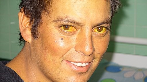
© Sheila J Toro, CC BY 4.0, via Wikimedia Commons
Investigations513
See the separate Abnormal liver function tests article.
Jaundice will be apparent if the total bilirubin is >35 μmol/L. It is usually easy to differentiate pre-hepatic causes of jaundice from hepatic and post-hepatic. It is more difficult to differentiate hepatic and post-hepatic, as they often co-exist (eg, obstructive jaundice with biliary cirrhosis).
In jaundice, the essential and rapid differentiation of the main causes (hepatitis, biliary stasis, haemolysis, resolution of haematoma or congenital causes) can often be achieved by performing the following investigations.
Levels of conjugated/unconjugated bilirubin (direct/indirect bilirubin).
Raised unconjugated (indirect) bilirubin suggests:
Gilbert's syndrome.
Haemolysis (reticulocytes, increased urinary urobilinogen, reduced serum haptoglobin).
Mild chronic hepatitis.
Crigler-Najjar syndrome (levels over 85 μmol/L).
Raised conjugated (direct) bilirubin (>10 μmol/L) suggests obstructive jaundice, including:
Liver disease
Pancreatic disease
Dubin-Johnson syndrome
FBC should include a reticulocyte count and blood smear to detect haemolysis.
ESR may be elevated - for example, in primary biliary cholangitis.
Lactate dehydrogenase is raised in haemolysis.
LFTs:
Alkaline phosphatase: considerably increased with either extrahepatic or intrahepatic biliary disease. The most common diseases associated with raised alkaline phosphatase include:
Gallstones causing bile duct obstruction.
Pancreatic cancer.
Pregnancy.
Drugs.
More rarely, PBC.
(Remember: Alkaline phosphatase is commonly raised in children as it becomes elevated during bone growth).
Serum transaminases are usually very high in hepatocellular disease (like viral hepatitis) but more modestly elevated in chronic hepatocellular damage and obstruction:
Aspartate aminotransferase (AST) is raised more than alanine aminotransferase (ALT) in cirrhosis, intrahepatic neoplasia, haemolytic jaundice and alcoholic hepatitis.
ALT is raised more than AST in acute hepatitis and in extrahepatic obstruction.
ALT levels of less than 100 IU/L with jaundice suggest obstructive jaundice.
ALT over 400 IU/L suggests diffuse acute hepatocellular damage (for example, in viral hepatitis).
ALT between 150-400 IU/L suggests chronic active hepatitis or viral or drug-induced hepatitis.
Very high levels of ALT (over 1,000 IU/L) suggest acute parenchymal disease.
Gamma-glutamyl transferase (GGT):
GGT is sensitive but not specific for excess alcohol intake.
A raised MCV with raised GGT is suggestive of alcohol abuse and, if accompanied by raised ALT, suggests liver cell damage.
Biliary obstruction and hepatic malignancies cause very high GGT levels (x 10 normal).
Raised GGT with raised alkaline phosphatase (over x 3 normal) suggests cholestasis.
Hepatitis serology should be done in all patients with cholestasis, as differentiating hepatitis from extrahepatic obstructive causes may be very difficult.
Prothrombin time may be prolonged because of vitamin K malabsorption. Injection of vitamin K will correct deficiency in cholestasis but not in parenchymal liver disease.
Serum antinuclear antibodies (ANAs), anti-smooth muscle antibody (ASMA): the hallmark of primary biliary cholangitis is antimitochondrial antibodies (90-95% of patients with primary biliary cholangitis are positive); ANA is positive in 20-50% of patients with primary biliary cholangitis.
Serum immunoglobulins and serum electrophoresis: IgG is raised in acute hepatitis, IgM is raised in autoimmune disease, primary biliary cholangitis or chronic infection.
Alpha-1-antitrypsin levels: deficiency causes cirrhosis and emphysema.
Ferritin to screen for haemochromatosis.
Imaging:
Abdominal ultrasound can detect liver abnormalities, hepatosplenomegaly and gallstones. It is useful to identify the extrahepatic causes of biliary obstruction but is also good at identifying intrahepatic disease (for example, malignant disease).14
Endoscopic ultrasound is commonly used to image the pancreas and biliary tract.15
CT scan.
ERCP is accurate at diagnosing benign and extrahepatic obstruction and can be combined with procedures to relieve obstruction.16
MRI scanning and magnetic resonance cholangiopancreatography (MRCP) provides images which are diagnostically-equivalent images to ERCP and is a useful technique in high risk patients to avoid significant morbidity. 1718
Liver biopsy can be done laparoscopically or percutaneously. It may be necessary, for example, to stage disease in PBC.
Jaundice treatment and management
This will depend on the diagnosis and cause of the jaundice in each individual case but factors to consider here include managing any pruritus that may be present - including lifestyle and dietary advice and cholestyramine treatment.
Exclusive updates for healthcare professionals
Stay informed with the latest clinical updates, professional insights, and evidence-based guidance. The Patient Pro newsletter curates essential content for healthcare professionals—delivered straight to your inbox.
By subscribing you accept our Privacy Policy. You can unsubscribe at any time. We never sell your data.
Further reading and references
- Chapter 25 - The Acutely Jaundiced Patient; L L Wong and D Gleeson; Case Studies in Adult Intensive Care Medicine
- Jaundice in primary care: a cohort study of adults aged >45 years using electronic medical records; A Taylor et al; Family Practice
- Epidemiological and Diagnostic Profile of Jaundice in Adults at Gabriel Touré University Hospital, Mali; K Doumbia; Asian Journal of Research and Reports in Hepatology
- Jaundice in Adult in-Patients at a Tertiary General Hospital; J N Hung et al; Journal of Biosciences and Medicines
- Epidemiological profile, referral routes and diagnostic accuracy of cases of acute cholangitis among individuals with obstructive jaundice admitted to a tertiary-level university hospital: a cross-sectional study; P F da Costa Soares et al; Sao Paulo Medical Journal
- Joseph A, Samant H; Hyperbilirubinemia.
- Jaundice in the Emergency Department: Meeting the Challenges of Diagnosis and Treatment; Emergency Medicine Practice
- Pappas G, Christou L, Akritidis NK, et al; Jaundice of unknown origin: Remember zoonoses! Scand J Gastroenterol. 2006 Apr;41(4):505-8.
- Crum NF; Epstein Barr virus hepatitis: case series and review. South Med J. 2006 May;99(5):544-7.
- Hinson JA, Roberts DW, James LP; Mechanisms of acetaminophen-induced liver necrosis. Handb Exp Pharmacol. 2010;(196):369-405. doi: 10.1007/978-3-642-00663-0_12.
- Suppiah A, Perry EP; Jaundice as a presentation of phenol induced hepatotoxocity following injection sclerotherapy for haemorrhoids. Surgeon. 2005 Feb;3(1):43-4. doi: 10.1016/s1479-666x(05)80011-3.
- Fang CL, Chu JS, Hsieh MC, et al; Signet-ring cell carcinoma of the ampulla of Vater. J Formos Med Assoc. 2004 Oct;103(10):793-6.
- Worthington J, Chapman R; Primary sclerosing cholangitis. Orphanet J Rare Dis. 2006 Oct 24;1(1):41.
- Gondal B, Aronsohn A; A Systematic Approach to Patients with Jaundice. Semin Intervent Radiol. 2016 Dec;33(4):253-258. doi: 10.1055/s-0036-1592331.
- Gerstenmaier JF, Gibson RN; Ultrasound in chronic liver disease. Insights Imaging. 2014 Aug;5(4):441-55. doi: 10.1007/s13244-014-0336-2. Epub 2014 May 24.
- Endoscopic ultrasound; D J Bell; Radiopaedia
- Tse F, Barkun JS, Romagnuolo J, et al; Nonoperative imaging techniques in suspected biliary tract obstruction. HPB (Oxford). 2006;8(6):409-25. doi: 10.1080/13651820600746867.
- Magnetic resonance cholangiopancreatography (MRCP); A Er; Radiopaedia
- Evaluation of Jaundice in Adults; M Nelson et al; American Family Physician
About the authorView full bio

Dr Philippa Vincent, MRCGP
General Practitioner, Medical Author
MB BS, Bsc, MRCGP (2000), DCH, DFSRH, DRCOG
Dr Philippa Vincent is an NHS GP working in North London.
About the reviewerView full bio

Dr Toni Hazell, FRCGP
MBBS, BSc, FRCGP, DFSRH, Dip GU med, DRCOG, DCH (London, UK, 2000)
Dr. Toni Hazell qualified from St. Mary’s Hospital Medical School and did her VTS at Northwick Park Hospital.
Article history
The information on this page is written and peer reviewed by qualified clinicians.
Article also available in English, German, Spanish, French, Italian, Portuguese, Hindi, Hebrew, Arabic, and Swedish.
Next review due: 29 Jan 2031
30 Jul 2026 | Latest version

Ask, share, connect.
Browse discussions, ask questions, and share experiences across hundreds of health topics.

Feeling unwell?
Assess your symptoms online for free
More in history and examination
- Chronic cough in adults
- Death certification
- Flashes, floaters and haloes
- Genitourinary history and examination - female
- Hiccups
- History and physical examination
- Nightmare disorder
- Nystagmus
- Obesity in children
- Palmar erythema
- Post-inflammatory hyperpigmentation of skin
- Post-traumatic stress disorder
- Presbyacusis
- Prevention of falls in elderly people
- Proctalgia fugax and anal pain
- Psychiatric assessment
- Stokes-Adams attacks
- Superficial thrombophlebitis
- Supraventricular tachycardia in adults