Cataracts and cataract surgery
Peer reviewed by Dr Philippa Vincent, MRCGPLast updated by Dr Toni Hazell, FRCGPLast updated 29 Jul 2026
Meets Patient’s editorial guidelines
- DownloadDownload
- Share
- Language
- Discussion
- Audio Version
- Add to preferred sources on Google
Medical Professionals
Professional Reference articles are designed for health professionals to use. They are written by UK doctors and based on research evidence, UK and European Guidelines. You may find the Cataracts article more useful, or one of our other health articles.
What are cataracts?
Cataracts are lens opacities. Some are small and do not need treatment but they often become large enough to block light and obstruct vision.
What causes cataracts?1
Ageing is the main cause of cataract development in the developed world, although they can also form congenitally and after various forms of injury to the lens. In the developing world other factors contribute, including malnutrition, acute dehydrating illnesses and excess ultraviolet (UV) exposure. Lack of access to treatment makes them a leading cause of sight loss worldwide.
How common are cataracts? (Epidemiology)2
The prevalence of cataract increases with age: a systematic review found the pooled prevalence (the prevalence of members of samples pooled together) of any cataract was:
20-39 years - 3.01%.
40-59 years - 16.97%
Over 60 years - 54.38%.
A UK study that randomly sampled 1,547 people of 65 years of age and older found that the prevalence of visually-impairing cataracts was:
71% in people aged over 85 years.
59% in people aged 80-84 years.
42% in people aged 75-79 years.
24% in people aged 70-74 years.
16% in people aged 65-69 years.
Cataracts are uncommon in children.
Globally, cataracts are the world's leading cause of sight loss. About 36 million people worldwide have lost their sight, and in over 12 million of them this is due to cataract.
Risk factors of cataracts1
Apart from age, the most significant types of cataract risk factors in the developed world are:
Smoking.
Diabetes mellitus.
Systemic corticosteroids.
Other risk factors contributing to cataract formation include:
Eye trauma.
Female gender.
Uveitis.
UV exposure.
Poor nutrition.
Lower socio-economic status.
Alcohol.
Toxins - eg, drugs of misuse.
Dehydrating illness crises.
Metabolic disorders - eg, galactosaemia in children.
Inflammatory and degenerative eye diseases.
Genetic studies estimate that the heritability of an age-related cataract is around 50%.
Evidence that topical (including inhaled) steroids alone increase the risk of cataract formation is unclear, but there is some evidence to suggest an increased risk from long-term use of steroid eye drops and topical steroids on the peri-orbital skin.3 4
In the developing world the main extrinsic factors are:
Diet (malnutrition).
Acute dehydrating diseases.
Cumulative exposure to sunlight.
In developing countries, cataracts are common in young adults and may be associated with atopy and with diabetes.
Can cataracts be improved with diet?5
The development of acquired cataract is principally caused by oxidative damage, which naturally leads to consideration of ameliorating this damage by increasing the amount of antioxidants in the diet. However, no strategy has provided convincing evidence of effectiveness in slowing cataract growth, although a 2023 review suggested that the incidence may be less in those who eat more plant-based products.6
Future research needs to take into account the underlying physiology of how specific nutrients and antioxidants are delivered, taken up and metabolised to maintain and restore antioxidant levels in the different regions of the lens.
Pathophysiology of cataract7 8
The lens continues to grow after birth, with the new secondary fibres being added as outer layers. New lens fibres are generated from the lens epithelium. Old fibres are not removed.
Lens transparency is maintained by many factors, including its microscopic structure and chemistry. The lens has three main parts: the capsule, the epithelium and the fibres. The lens has no nerves, blood vessels, or connective tissue.
The lens capsule is elastic and is composed of collagen.
The lens epithelium, in the anterior portion of the lens between the capsule and the fibres, regulates the homeostasis of the lens and constantly lays down new fibres.
The lens fibres form the bulk of the lens. They are long, thin, transparent cells, arranged lengthwise from the posterior to the anterior poles. They are stacked arranged in concentric layers. They have no nuclei and are composed mainly of crystallins (water-soluble proteins). The transparency of the lens is maintained by the arrangements of these fibres, their lack of intracellular bodies like nuclei, and their cytoskeletons which maintain the architecture.
Transparency is maintained by the structure of the lens proteins and by the way they are stacked, linked and aligned. Disruption of the crystallin fibres will affect the integrity of the carefully composed structure, leading to protein aggregation. Cataracts result from the deposition of aggregated proteins in the lens, causing clouding, light scattering, double vision and obstruction of vision.
A second contributing factor which occurs with ageing is an accumulation of yellow-brown pigment in the lens. This does not affect image sharpness but it affects colour vision and contrast, so may eventually make reading difficult.9
Cataract symptoms10
What are the symptoms of cataracts?
This depends upon the size and location of the opacity and whether one or both eyes are affected. The most consistent presenting features are:
Gradual painless loss of vision.
Difficulties with reading.
Failure to recognise faces.
Problems watching TV.
Diplopia in one eye.
Haloes.
Many cataracts present before they are symptomatic, because they are noticed by an optician at a routine eye exam.
Cataracts are often described by the part of the lens that is particularly affected. This also affects their typical presentation. There are many possible subdivisions but three broad categories are generally described:
Nuclear sclerosis: this cataract is formed by new layers of fibre (added with ageing) compressing the nucleus of the lens. Typical symptoms include:
Gradually reduced contrast.
Reduced colour intensity.
Reading which may be surprisingly good for acuity level.
Difficulty in seeing golf balls, car number plates, etc.
Difficulty in recognising faces.
Cortical: new fibres are added to the outside of the lens, which age and produce cortical spokes. These may not produce symptoms unless on the visual axis or the entire cortex is affected when it is 'mature':
Light scatter from opacities.
Problems with glare when driving, particularly at night.
Difficulty reading.
Daytime activity relatively unaffected as the iris is constricted.
Posterior subcapsular: opacities in the central posterior cortex. This may occur in younger patients and may cause glare ± deterioration in near vision:
Visually disabling in good lighting - less trouble at low light levels when the pupil is dilated.
Difficulty in daytime driving.
Difficulty in reading.
Paediatric cataracts2
These may be:
Congenital : hereditary/genetic, metabolic (eg, galactosaemia), in-utero infection (toxoplasmosis, other, rubella, cytomegalovirus, herpes simplex (TORCH)).
Developmental: genetic, metabolic (eg, galactokinase deficiency).
Acquired: metabolic (eg, diabetes mellitus), traumatic, post-radiotherapy.
The most common cause of congenital cataracts is infection - rubella (the most common), measles, chickenpox, cytomegalovirus, herpes simplex, herpes zoster, poliomyelitis, influenza, Epstein-Barr virus, syphilis and toxoplasmosis. Other causes are metabolic and genetic syndromes.
Signs of cataracts
Opacities can be seen as defects in the red reflex obtained when the ophthalmoscope is held 60 cm from the eye. This is best seen with a dilated pupil.
The lens may appear brown or white when a bright light is shone on the eye.
Check that:
Visual acuity is not improved by viewing test through a pinhole.
The patient can indicate where a light is placed.
Pupillary reactions are normal.
Differential diagnosis
Cataract treatment and management1
There is no proven prevention or medical treatment for a cataract. Modern cataract surgery involves lens extraction and replacement. The technique can be intracapsular or extracapsular, although intracapsular extraction is now rarely performed. Surgical removal of the cataract is the only effective treatment to restore or maintain vision.
In the developed world, cataracts are easily treated before vision is severely impaired. In the developing world, cataracts lead to significant problems of severe visual impairment. This is because most people do not seek advice until the cataract is advanced or the eye has developed lens-induced glaucoma, a painful condition. This is partly due to lack of awareness and partly due to a lack of government or socially funded healthcare in countries where socio-economic conditions are poor. Many countries lack sufficient clinicians to meet demand, particularly in rural areas.
Cataract surgery210
There is no absolute threshold of visual acuity at which surgery is indicated. The National Institute for Health and Care Excellence (NICE) advises that the decision to refer a person with a cataract for surgery should be based on a discussion with them that includes: how the cataract affects the person's vision and quality of life; whether one or both eyes are affected; what cataract surgery involves, including possible risks and benefits; how the person's quality of life may be affected if they choose not to have cataract surgery and whether the person wants to have cataract surgery. It states that access to cataract surgery should not be restricted on the basis of visual acuity.
Cataract
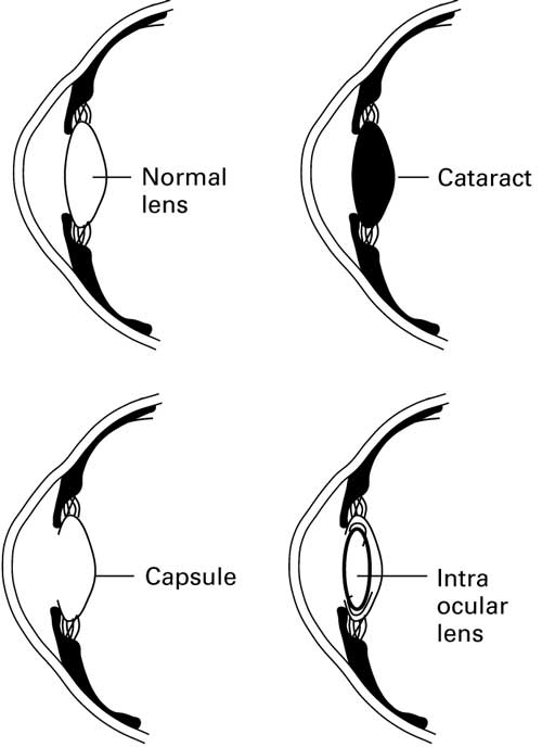
Complications of cataract surgery1
Cataract surgery is a safe and successful procedure in the majority of cases. Poor vision after cataract surgery is generally due to:
Inadequate correction of refractive error postoperatively.
Failure to detect pre-existing eye conditions. Serious co-existing eye conditions such as glaucoma, age-related macular degeneration, diabetic retinopathy and amblyopia are often present in patients requiring cataract surgery.
Surgical complications.
The most common complication is capsular rupture with vitreous loss, which is significant, as it is often associated with a poorer outcome. It also increases the risk of endophthalmitis, which is rare but devastating.
Early complications of cataract surgery
Posterior capsule rupture with or without loss of vitreous (1-3%).
Protruding or broken sutures.
Trauma to the iris.
Wound gape or prolapse of iris (<1%).
Anterior chamber haemorrhage (<1%).
Vitreous haemorrhage (<1%).
Choroidal haemorrhage (<1%).
Endophthalmitis - a devastating but rare condition which occurs in less than 0.5% of cases. Staphylococcus epidermidis is the most common infecting organism.
Refractory uveitis due to low-grade infection. This can be avoided by intraoperative antibiotic prophylaxis.
Late complications of cataract surgery1
Posterior capsule opacification - this is a late complication (around 40%) and is the most common finding. Vision can usually be restored with laser capsulotomy.
Cystoid macular oedema, more frequently diagnosed by angiography (1%).
Retinal detachment, which can occur weeks, months or even years later. The lifetime risk is about 1 per 150 operations, more likely in case of high myopia.
Open-angle glaucoma (<1%).
Closed-angle glaucoma (<1%).
Bullous keratopathy (a rare complication).
Dysphotopsias:
Negative and positive dysphotopsias are unwanted optical phenomena occurring after cataract surgery due to intraocular light artefacts reflecting off the IOL.
They are the main cause of post-surgical dissatisfaction and are seen in up to two-thirds of patients immediately after cataract surgery, but only around 2-3% after a year. 11
Positive dysphotopsia (glare, haloes and starbursts) is largely attributed to edge effects of the implant, whilst negative dysphotopsia appears related to the patient's anatomical structure.
There are no comprehensive figures on outcomes of cataract surgery in developing countries and on the relative importance of spectacles, patient selection and surgery. However, at least 25% of the six million cataract operations performed annually in developing countries are believed to have poor outcomes. About one quarter of these are due to surgical complications.12
Prognosis12
Adults
Age-related cataracts are progressive and the progression is variable and unpredictable. Without treatment, most people with a cataract will develop severe visual impairment.
The visual performance of the eye is generally considered in terms of acuity, contrast sensitivity, glare disability, visual fields, and colour vision. Outcomes for cataract surgery have mainly been assessed in terms of acuity only.
The 2024 National Ophthalmology Database Audit found that the risk of severe complications continues to fall - to 0.79% for posterior capsule rupture, 0.48% for vision loss and 0.21% for severe vision loss. 13
With surgery, 95% of people will have 6/12 best corrected vision if there is nothing else wrong with the eye.
More recent studies have looked at self-reporting of outcomes and these make it clear that patients may consider surgery unsuccessful in the face of improved visual acuity if, for example, anisometropia or disturbance from the fellow eye results.
Pre-operative visually significant ocular comorbidity was the most important predictor of poor subjective outcome. Older age was not per se. The greatest benefit was seen for second eye surgery in younger patients.
Patients may be disappointed to find that they require glasses for distant vision after cataract surgery, when they did not require them prior to surgery.
Children
Untreated cataracts in children younger than 10 years of age cause amblyopia, leading to lifelong visual impairment even if the cataracts are later removed.
Most children with a unilateral cataract have normal vision in the eye without the cataract. Most children who have treatment for bilateral cataracts achieve only partial sight.
Driving14
Advise the patient not to drive and to contact the DVLA if they cannot meet the criteria listed below. That is likely to be the case where there are severe bilateral cataracts, or after failed bilateral cataract extractions:
Group 1 entitlement (to drive a car or motorcycle)
To read in good daylight (with the aid of glasses or contact lenses if worn) a registration mark fixed to a motor vehicle and containing letters and figures 79 millimetres high and 50 millimetres wide at a distance of 20 metres, or at a distance of 20.5 metres where the characters are 79 millimetres high and 57 millimetres wide.
Visual acuity (with the aid of glasses or contact lenses if needed) must be at least 6/12 with both eyes open (or in the only eye if you have vision in one eye only).
Note that in the presence of cataract, glare may affect your ability to meet the number plate requirements, even if your acuity is good enough.
Group 2 entitlement (to drive a larger vehicle)
To have a visual acuity, using corrective lenses if necessary, of at least 6/7.5 (0.8 decimal) in the better eye and at least 6/12 (0.5 decimal) in the other eye.
The uncorrected acuity in each eye must be at least 3/60.
Where glasses are worn to meet the minimum standards, they should have a corrective power ≤+8 dioptres.
It is also necessary for all drivers of Group 2 vehicles to be able to meet the prescribed and relevant Group 1 visual acuity requirements.
In the presence of a cataract, glare may affect the ability to meet the number plate requirement, even with appropriate acuities.
If there is any uncertainty about fitness to drive, advise the patient to contact the DVLA or to seek clarification from an eye specialist.
Dr Mary Lowth is an author or the original author of this leaflet.
Exclusive updates for healthcare professionals
Stay informed with the latest clinical updates, professional insights, and evidence-based guidance. The Patient Pro newsletter curates essential content for healthcare professionals—delivered straight to your inbox.
By subscribing you accept our Privacy Policy. You can unsubscribe at any time. We never sell your data.
Further reading and references
- Nizami AA, Gulani AC; Cataract
- Cataracts; NICE CKS, March 2025 (UK access only)
- Daniel BS, Orchard D; Ocular side-effects of topical corticosteroids: what a dermatologist needs to know. Australas J Dermatol. 2015 Aug;56(3):164-9. doi: 10.1111/ajd.12292. Epub 2015 Mar 5.
- Kramer M, Tomkins-Netzer O; Cataract Risk and Topical Corticosteroids among Children with Juvenile Idiopathic Arthritis-Related Uveitis. Ophthalmology. 2020 Apr;127(4S):S19-S20. doi: 10.1016/j.ophtha.2019.10.042.
- Braakhuis AJ, Donaldson CI, Lim JC, et al; Nutritional Strategies to Prevent Lens Cataract: Current Status and Future Strategies. Nutrients. 2019 May 27;11(5). pii: nu11051186. doi: 10.3390/nu11051186.
- Falkowska M, Mlynarczyk M, Micun Z, et al; Influence of Diet, Dietary Products and Vitamins on Age-Related Cataract Incidence: A Systematic Review. Nutrients. 2023 Oct 28;15(21):4585. doi: 10.3390/nu15214585.
- Zhao L, Chen XJ, Zhu J, et al; Lanosterol reverses protein aggregation in cataracts. Nature. 2015 Jul 30;523(7562):607-11. doi: 10.1038/nature14650. Epub 2015 Jul 22.
- Andley UP; Effects of alpha-crystallin on lens cell function and cataract pathology. Curr Mol Med. 2009 Sep;9(7):887-92.
- Allen D, Vasavada A; Cataract and surgery for cataract. BMJ. 2006 Jul 15;333(7559):128-32. doi: 10.1136/bmj.333.7559.128.
- Cataracts in adults: management; NICE Guideline (Nov 2017)
- Pusnik A, Petrovski G, Lumi X; Dysphotopsias or Unwanted Visual Phenomena after Cataract Surgery. Life (Basel). 2022 Dec 24;13(1):53. doi: 10.3390/life13010053.
- Yorston D; Cataract complications. Community Eye Health. 2008 Mar;21(65):1-3.
- Latest audit figures show increase in numbers and outcomes of cataract procedures; Royal College of Ophthalmologists, May 2024
- Assessing fitness to drive: guide for medical professionals; Driver and Vehicle Licensing Agency
About the authorView full bio

Dr Toni Hazell, FRCGP
MBBS, BSc, FRCGP, DFSRH, Dip GU med, DRCOG, DCH (London, UK, 2000)
Dr. Toni Hazell qualified from St. Mary’s Hospital Medical School and did her VTS at Northwick Park Hospital.
About the reviewerView full bio

Dr Philippa Vincent, MRCGP
General Practitioner, Medical Author
MB BS, Bsc, MRCGP (2000), DCH, DFSRH, DRCOG
Dr Philippa Vincent is an NHS GP working in North London.
Article history
The information on this page is written and peer reviewed by qualified clinicians.
Article also available in English, German, Spanish, French, Italian, Portuguese, Hindi, Hebrew, Arabic, and Swedish.
Next review due: 29 Jan 2031
29 Jul 2026 | Latest version

Ask, share, connect.
Browse discussions, ask questions, and share experiences across hundreds of health topics.

Feeling unwell?
Assess your symptoms online for free
More in ophthalmology
- Chalazion
- Chorioretinal inflammation
- Congenital nasolacrimal duct obstruction
- Contact lenses
- Corneal abrasions, foreign bodies and injuries
- Diabetic retinopathy and diabetic eye problems
- Eye drugs - prescribing and administering
- Eye injuries (assessment, treatment and management)
- Förster-Fuchs retinal spot
- Horner's syndrome
- Infective conjunctivitis
- Inflammatory retinal diseases
- Keratomalacia
- UpdatedOptic disc swelling
- Posterior vitreous detachment
- Red eye
- Retinal tumours
- Severe and partial sight impairment
- Subconjunctival haemorrhage
- Tumours of the choroid