DMSA-skanning
Granskad av Dr Toni Hazell, MRCGPSenast uppdaterad av Dr Doug McKechnie, MRCGPSenast uppdaterad 14 Nov 2023
Uppfyller patientens redaktionella riktlinjer
- Ladda nerLadda ner
- Dela
- Language
- Diskussion
- Ljudversion
- Lägg till i föredragna källor på Google
A DMSA scan uses a radioactive chemical to create specialised pictures of the kidneys. It can help to show whether the kidneys are damaged or scarred.
Notera: informationen nedan är endast en allmän vägledning. Arrangemangen och sättet tester utförs på kan variera mellan olika sjukhus. Följ alltid instruktionerna från din läkare eller lokala sjukhus.
Överblick
A DMSA scan uses radioactive chemicals to take pictures of your kidneys.
It helps doctors assess kidney function, shape, and identify scarring.
The scan is often used for children with unusual or repeated urinary tract infections.
A small amount of radioactive substance is injected and builds up in the kidneys.
You will need to wait a few hours between the injection and the scan itself.
Inform the hospital if you are pregnant, may be pregnant, or are breastfeeding.
After the scan, drink plenty of fluids to help flush out the radioactive chemical.
I den här artikeln:
Videoval för Bilddiagnostik
Fortsätt läsa nedan
What is a DMSA scan?
DMSA stands for dimercaptosuccinic acid. A DMSA scan uses radioactive chemicals to create special pictures of the kidneys. An example of a DMSA scan of the kidneys is below.
DMSA scan of the kidneys
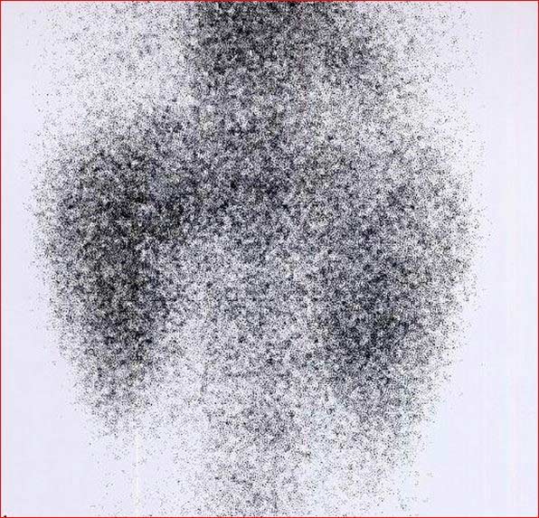
These pictures can help doctors assess how well the kidneys are working. DMSA travels through the body joined to a radioactive chemical. It builds up in the kidneys. Pictures of the kidneys are then taken using a special camera which can detect the radioactive chemical.
What is a DMSA scan used for?
Tillbaka till innehållThis scan is used to check the structure of the kidneys, their size and shape. It is often used in children who have had unusual or repeated urinary tract infections.
It shows which areas of the kidney are working well and any areas of scarring. Scarring can be caused by a condition in which urine travels back from the bladder to the kidneys. This is called vesico-ureteric reflux. DMSA scans can also look for damage following an injury or reduced blood supply to the kidneys.
A DMSA scan enables doctors to see the functioning tissue of your kidneys. This is because DMSA does not attach itself to areas of the kidneys that are damaged. Doctors can compare the function of each kidney to see if one kidney functions differently to the other. By performing regular DMSA scans they can monitor any changes to inflammation of the kidneys.
Other investigations such as an ultrasound scan can show the size and shape of the kidneys but not how they are working. This is why a DMSA scan may often be recommended in addition to other kidney tests or scans.
DMSA scans are usually requested by hospital specialists (usually paediatricians - doctors who specialise in children). The results should be given by the hospital specialist who requested the test, not by the GP.
Fortsätt läsa nedan
How does a DMSA scan work?
Tillbaka till innehållA DMSA scan is a type of radionuclide scan. A radionuclide (sometimes called a radioisotope or isotope) is a chemical which emits a type of radioactivity called gamma rays.
A tiny amount of radionuclide is put into the body, usually by an injection into a vein. The radioactive chemical is often coupled to another substance. This substance takes the radioactive chemical to the part of the body doctors want to see.
It is possible to make many different types of radionuclides. Different ones tend to build up or concentrate in different organs or tissues. So, the radionuclide used depends on which part of the body is to be scanned.
In this case the substance DMSA is used because it builds up in the kidneys. Cells which are most 'active' in the kidney will take up more of the DMSA. So, active parts of the kidney tissue will emit more gamma rays than less active or inactive parts.
Gamma rays are similar to X-rays and are detected by a device called a gamma camera. The gamma rays which are emitted from inside the body are detected by the gamma camera, are converted into an electrical signal, and sent to a computer.
The computer builds a picture by converting the differing intensities of radioactivity emitted into different colours or shades of grey. For example, areas of the target organ or tissue which emit lots of gamma rays may be shown as red spots ('hot spots') on the picture on the computer monitor.
Areas which emit low levels of gamma rays may be shown as blue ('cold spots'). Various other colours may be used for 'in between' levels of gamma rays emitted.
What happens during a DMSA scan?
Tillbaka till innehållA DMSA scan is usually done in the nuclear medicine department of a hospital. The first part of the scan involves a small injection. This is usually into a vein in the back of the hand. Children being scanned may be asked to come to the scanning unit an hour or so before the injection. This allows staff the time to put special Emla® cream on the back of a child's hand. Emla® cream is a local anaesthetic that numbs the area to reduce the discomfort of the injection.
You may also be asked to provide a urine sample (specimen) to make sure you do not have an active urine infection at the time of the test. If there is an infection, or if you have had a very recent infection, the scan may need to be postponed. This is because infection can alter the results of the scan and make it unreliable.
After the injection there will be a delay, usually around two to four hours, before the scan takes place. This allows enough time for the DMSA substance to travel around the body and reach the kidneys. Some hospitals will ask you to have some water or go to the toilet during this time, as this may make the pictures clearer.
After the wait the gamma camera is used to take pictures of the kidneys. It can take approximately 45 minutes to take all the pictures. During this time you need to remain as still as possible. If you are taking a child to be scanned it might be useful to bring your child's favourite book or toy to keep them occupied.
The camera can be quite big and for some pictures it may come quite close to your child's tummy (abdomen). It may be helpful to explain this before the test. Parents are usually allowed to stay with their children throughout the test.
Fortsätt läsa nedan
How to prepare for a DMSA scan
Tillbaka till innehållYour local hospital should give you information on how to prepare for the scan. If you are pregnant, think you may be pregnant or are breastfeeding you must let the hospital know before the scan. You may be asked to drink lots of fluids before you attend.
Some hospitals recommend that you avoid medicines containing certain substances before the test. Your doctor should be able to advise you on this.
What to expect after a DMSA scan
Tillbaka till innehållThe radioactive chemical you receive is eliminated from your body through the urine over the 24 hours following the scan. For that reason, you should drink plenty of fluids and urinate frequently following the injection. How much fluid will depend on each individual but you should be well hydrated and, for an adult, this could be 3-4 glasses of water.
The colour of your urine won't be affected by a DMSA scan. However, as it contains the radioactive tracer, it is recommended that you wash your hands well after going to the toilet.
In the case of babies and youngsters in nappies who are having a DMSA scan, there will be a small amount of radioactivity in the urine and therefore on the child's nappy. The radiotracer will not affect the baby's skin; however, carers should wash the baby's bottom as normal and wash their hands thoroughly.
Cloth nappies need to be washed thoroughly and disposable nappies put in a plastic bag and sealed before being disposed of.
If you have contact with children or pregnant women you should let your doctor know. Although the levels of radiation used in the scan are small the doctor may advise special precautions. Your hospital will be able to give you advice on this.
Possible side-effects or complications from a DMSA scan
Tillbaka till innehållThe term 'radioactivity' may sound alarming. But, the radioactive chemicals used in radionuclide scans are considered to be safe and they leave the body quickly in the urine.
The dose of ionising radiation that your body receives is very small. In many cases, the level of radiation involved is not much different to a series of a few normal X-rays. However:
As with any other types of radiation (such as X-ray), there is a small risk that the gamma rays may affect an unborn baby. So, tell your doctor if you are pregnant or if you may be pregnant.
The scan usually isn't done if you are pregnant, unless it's absolutely necessary and can't be delayed until after the pregnancy.
If you're pregnant and your child is having a DMSA scan, it's usually recommended to get another adult to help look after them for the first 24 hours after the scan, as a precaution - and to avoid any contact with your child's bodily fluids (wee, poo, and vomit) for that time.
Rarely, some people have an allergic reaction to the injected chemical.
Theoretically, it is possible to receive an overdose when the chemical is injected. This is very rare.
Understanding the kidneys
Tillbaka till innehållRenal tract
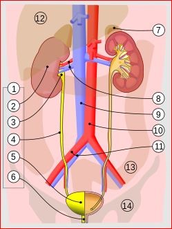
© Jordi March i Nogué [1], CC BY-SA 3.0, via Wikimedia Commons
There are two kidneys, one on each side of the tummy (abdomen). They make urine which drains down the ureters into the bladder. The ureters are tubes which go from each kidney to the bladder. Urine is stored in the bladder and, from time to time, is passed out through a tube called the urethra when we go to the toilet.
The kidneys, ureters, bladder and urethra are called the urinary tract.
Patientval för Bilddiagnostik

Tester och undersökningar
Benscanning
En skelettscintigrafi är en speciell typ av nukleärmedicinsk procedur som använder radionuklider för att skapa en bild av skelettet. Radionuklider är kemikalier som avger radioaktivitet som kan upptäckas av speciella skannrar. En skelettscintigrafi skiljer sig från en bentäthetsmätning (DEXA). Se broschyren om DEXA-skanning för mer information om detta bentäthetstest. En DEXA-skanning är också känd som en DXA-skanning. Obs: informationen nedan är endast en allmän vägledning. Arrangemangen och sättet tester utförs på kan variera mellan olika sjukhus. Följ alltid instruktionerna från din läkare eller lokala sjukhus.
av Dr Doug McKechnie, MRCGP

Tester och undersökningar
PET scan
A PET scan create images which show where cells are particularly active in the body. It is most commonly used to diagnose and assess cancer. Note: the information below is a general guide only. The arrangements, and the way tests are performed, may vary between different hospitals. Always follow the instructions given by your doctor or local hospital.
av Dr Rosalyn Adleman, MRCGP
Vanliga frågor
Why would doctors recommend a DMSA scan instead of just an ultrasound?
While an ultrasound scan can show the size and shape of the kidneys, it cannot assess how well they are working. A DMSA scan, however, shows which areas of the kidney tissue are functional and can detect scarring or damage, providing a more complete picture of kidney health.
How long after the injection is the scan actually performed?
There is a delay of about two to four hours between the injection and the actual scanning process. This waiting period allows sufficient time for the DMSA substance to travel through the body and accumulate in the kidneys, ensuring clear pictures.
Can I eat normally before a DMSA scan?
The article mentions that you may be asked to drink plenty of fluids before attending the scan. It also states that some hospitals recommend avoiding certain medications, but it does not specify any dietary restrictions regarding food. Your hospital should provide specific preparation instructions.
What should I do if my baby wears cloth nappies after a DMSA scan?
If your baby wears cloth nappies after a DMSA scan, these nappies should be washed thoroughly. Disposable nappies, on the other hand, should be placed in a plastic bag, sealed, and then disposed of as normal waste.
What is the likelihood of having an allergic reaction to the DMSA injection?
Allergic reactions to the injected chemical are rare. The article states that this happens only rarely.
Vidare läsning och referenser
- Shaikh N, Spingarn RB, Hum SW; Dimercaptosuccinic acid scan or ultrasound in screening for vesicoureteral reflux among children with urinary tract infections. Cochrane Database Syst Rev. 2016 Jul 5;7:CD010657. doi: 10.1002/14651858.CD010657.pub2.
- Urinary tract infection - children; NICE CKS, April 2024 (UK access only)
Fortsätt läsa nedan
Om författarenVisa fullständig biografi

Dr Doug McKechnie, MRCGP
Medicinsk skribent
MA, MBBS, MSc, DRCOG, MRCP(UK), MRCGP(2021), FHEA
Dr Doug McKechnie är en NHS-läkare som arbetar i London. Han arbetar kliniskt på heltid och är också biträdande ansvarig för modulen Klinisk och Professionell Praxis vid University College London Medical School.
Om recensentenVisa fullständig biografi

Dr Toni Hazell, MRCGP
MBBS, BSc, MRCGP, DFSRH, Dip GU med, DRCOG, DCH (London, UK, 2000)
Dr. Toni Hazell tog examen från St. Mary’s Hospital Medical School och genomförde sin VTS vid Northwick Park Hospital.
Artikelhistorik
Informationen på denna sida är skriven och granskad av kvalificerade kliniker.
Next review due: 12 Nov 2028
14 Nov 2023 | Senaste versionen

Fråga, dela, anslut.
Bläddra i diskussioner, ställ frågor och dela erfarenheter inom hundratals hälsorelaterade ämnen.

Känner du dig sjuk?
Bedöm dina symtom online gratis
Anmäl dig till Patientens nyhetsbrev
Din veckovisa dos av tydliga, pålitliga hälsoråd - skrivna för att hjälpa dig känna dig informerad, självsäker och i kontroll.
Genom att prenumerera accepterar du våra Sekretesspolicy. Du kan avsluta prenumerationen när som helst. Vi säljer aldrig dina uppgifter.