Chondrodermatitis nodularis
Granskad av Dr Hayley Willacy, FRCGP Senast uppdaterad av Dr Colin Tidy, MRCGPSenast uppdaterad 10 feb 2023
Uppfyller patientens redaktionella riktlinjer
- Ladda nerLadda ner
- Dela
- Language
- Diskussion
- Ask Patient
- Ljudversion
- Lägg till i föredragna källor på Google
Medicinska yrkesverksamma
Professionella referensartiklar är utformade för att användas av hälso- och sjukvårdspersonal. De är skrivna av brittiska läkare och baserade på forskningsbevis, brittiska och europeiska riktlinjer. Du kanske hittar en av våra hälsoartiklar mer användbar.
Synonyms: chondrodermatitis nodularis helicis (or antihelicis - depending on the site of the lesion); chondrodermatitis nodularis chronica helicis; ear pressure sore; painful nodule of the ear; Winkler's disease
This is a painful, inflamed nodule of the ear, involving the cartilage and skin of the pinna. It is usually located at the apex of the helix.
Etiologi1
It is probably caused mainly by pressure, combined with the anatomy of the pinna and its blood supply:
The initial damage may be triggered by inflammation, oedema and necrosis from trauma, cold, actinic damage or pressure.
The pinna has relatively little subcutaneous tissue for insulation and padding. Only small dermal blood vessels supply the epidermis, dermis, perichondrium and cartilage. These features may prevent adequate healing and lead to secondary perichondritis.
Possibly, perichondrial arteriolar changes may be involved. An autoimmune aetiology has also been implicated.2
Epidemiologi3
The condition is probably relatively common but is rarely documented in the literature. It is most common in elderly men but It affects those of all ages, gender and pigmentation.
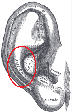
Data suggest that the condition is likely to be localised to the helix in men and the antihelix (the curved raised ridge of cartilage in front of the helix) in women.
Anything that puts pressure on the external ear, such as headphones, may precipitate the condition, as may a tendency always to sleep on the same side.
Antihelix position
Presentation4
History:
It presents as a painful nodule on the helix or antihelix of the ear.
It appears spontaneously and then usually enlarges rapidly to its maximum size and remains stable. Onset may be precipitated by pressure, trauma or cold.
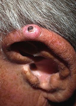
Chondrodermatitis nodularis of the ear
Skyltar
There is usually only one nodule - most often on the outer side of the helix, commonly at its apex:
The nodule is firm, tender and well demarcated.
Size range is 3-20 mm in diameter - usually about 6 mm in diameter.
It is round or oval, slightly raised and firmly stuck to the underlying cartilage.
It may appear inflamed.
The surface appears scaly or crusty. If the crust is removed, a small raw area or core may be visible centrally.
Differentialdiagnos5
Elastotic nodules of the ears.
Cystic chondromalacia.
Utredningar6
Biopsy is needed if the diagnosis is uncertain, particularly to exclude basal cell carcinoma or squamous cell carcinoma.
Associerade sjukdomar
The condition is benign. One study suggested an association with immune-mediated vascular compromise and necrobiotic collagenous disease such as granuloma annulare, especially in younger patients.7
Another report concerns a patient with systemic sclerosis and primary biliary cirrhosis (Reynold's syndrome). Such patients have an increased risk of developing skin malignancies.2
Ledning4
The lesion rarely resolves spontaneously and so will usually need some form of treatment if it is causing discomfort or annoyance. A trial of conservative therapy for all patients is suggested.1 8
Conservative management
The aim of treatment is to reduce pressure on the site of the lesion - for example, by:
Altering the way headphones or telephone receivers are held to the ear.
Sleeping on the opposite side, if possible.
Using a soft pillow, or modifying the pillow with a hole where the nodule is in contact. A doughnut-shaped pillow was effective in one study.8 A haemorrhoid cushion is a novel cheap alternative.9
Using a corn plaster.
A pressure-relieving device, which can be made by using foam plastic to be applied at night.7
Using a protective made-to-measure silicone splint.1
In addition:
Avoid excessive exposure to the cold and the sun.
Topical antibiotics for secondary infections or ulceration. Petroleum jelly may also help if the lesion is ulcerated.1
Further management10
Topical nitroglycerin has been beneficial in the control of symptoms and in improving appearance.11
Topical and intralesional steroids may relieve discomfort.
Cryotherapy has been used.
CO2 and argon lasers have been used with benefit in a small number of patients.
Photodynamic therapy has shown some promise and is currently being investigated.
Kirurgi:
There are various surgical techniques used to achieve excision of the affected cartilage with reconstruction of the overlying skin.
Excision of the damaged area of cartilage is usually successful but recurrence can occur at the edge of the excised area.
Narrow elliptical excision of the skin lesion with shaving of the underlying cartilage appears to be as effective and less invasive, and to have similar recurrence rates (around 13.5% after four years or so).12
Excision by punch biopsy, with full-thickness skin grafting (the 'punch and graft' technique).1
Prognos
Exklusiva uppdateringar för vårdpersonal
Håll dig informerad med de senaste kliniska uppdateringarna, professionella insikter och evidensbaserad vägledning. Patient Pro-nyhetsbrevet sammanställer viktigt innehåll för vårdpersonal—levererat direkt till din inkorg.
Genom att prenumerera accepterar du våra Sekretesspolicy. Du kan avsluta prenumerationen när som helst. Vi säljer aldrig dina uppgifter.
Vidare läsning och referenser
- Elsensohn A, Getty S, Shiu J, et al; Intradermal Proliferative Fasciitis Occurring With Chondrodermatitis Nodularis Helicis. Am J Dermatopathol. 2017 Nov 21. doi: 10.1097/DAD.0000000000001027.
- Kumar P, Barkat R; Chondrodermatitis nodularis chronica helicis. Indian Dermatol Online J. 2017 Jan-Feb;8(1):48-49. doi: 10.4103/2229-5178.198767.
- Chondrodermatitis nodularis helicis; DermNet NZ
- Sifuentes Giraldo WA, Gonzalez-Garcia C, de Las Heras Alonso E, et al; Chondrodermatitis nodularis chronica helicis in a patient with systemic sclerosis associated with primary biliary cirrhosis (Reynolds syndrome): A case report. Eur J Rheumatol. 2014 Dec;1(4):161-163. doi: 10.5152/eurjrheumatol.2014.140048. Epub 2014 Dec 1.
- Orengo I, Robbins K, Marsch A; Pathology of the ear. Semin Plast Surg. 2011 Nov;25(4):279-87. doi: 10.1055/s-0031-1288920.
- Salah H, Urso B, Khachemoune A; Review of the Etiopathogenesis and Management Options of Chondrodermatitis Nodularis Chronica Helicis. Cureus. 2018 Mar 26;10(3):e2367. doi: 10.7759/cureus.2367.
- Wagner G, Liefeith J, Sachse MM; Clinical appearance, differential diagnoses and therapeutical options of chondrodermatitis nodularis chronica helicis Winkler. J Dtsch Dermatol Ges. 2011 Apr;9(4):287-91. doi: 10.1111/j.1610-0387.2011.07601.x. Epub 2011 Jan 31.
- Chondrodermatitis nodularis helicis; Primärvårdens Dermatologiska Sällskap (PCDS)
- Kuen-Spiegl M, Ratzinger G, Sepp N, et al; Chondrodermatitis nodularis chronica helicis - a conservative therapeutic approach by decompression. J Dtsch Dermatol Ges. 2011 Apr;9(4):292-6. doi: 10.1111/j.1610-0387.2010.07572.x. Epub 2010 Nov 23.
- Sanu A, Koppana R, Snow DG; Management of chondrodermatitis nodularis chronica helicis using a 'doughnut pillow'. J Laryngol Otol. 2007 Nov;121(11):1096-8. Epub 2007 May 24.
- Ali FR, Healy C, Mallipeddi R; Hemorrhoid cushions for chondrodermatitis nodularis helicis (CNH): Piling off the pressure. J Am Acad Dermatol. 2016 Aug;75(2):e65-6. doi: 10.1016/j.jaad.2016.03.014.
- Juul Nielsen L, Holkmann Olsen C, Lock-Andersen J; Therapeutic Options of Chondrodermatitis Nodularis Helicis. Plast Surg Int. 2016;2016:4340168. doi: 10.1155/2016/4340168. Epub 2016 Jan 27.
- Flynn V, Chisholm C, Grimwood R; Topical nitroglycerin: a promising treatment option for chondrodermatitis nodularis helicis. J Am Acad Dermatol. 2011 Sep;65(3):531-6. doi: 10.1016/j.jaad.2010.06.012. Epub 2011 May 6.
- Rex J, Ribera M, Bielsa I, et al; Narrow elliptical skin excision and cartilage shaving for treatment of chondrodermatitis nodularis. Dermatol Surg. 2006 Mar;32(3):400-4.
Om författarenVisa fullständig biografi
Uppdaterad av

Dr Colin Tidy, MRCGP
Allmänläkare, Medicinsk Författare
MBBS, MRCGP, MRCP (Paediatrics), DCH
Dr Colin Tidy är en NHS-läkare, baserad i Oxfordshire.
Om recensentenVisa fullständig biografi

Dr Hayley Willacy, FRCGP
Allmänläkare, Medicinsk Författare
MBChB (1992), DRCOG, DFFP, MRCOG (Part 1) MRCGP (2007), DFSRH (2013), MSc - medical education (2020)
Dr Hayley Willacy var en NHS-läkare som arbetade i nordvästra England och gick i pension från klinisk praktik 2022 efter 30 år.
Artikelhistorik
Artikeln finns också på Engelska, Tyska, Spanska, Franska, Italienska, Portugisiska, Hindi, Hebreiska, Arabiska, och Svenska.
Nästa granskning: 9 feb 2028
10 feb 2023 | Senaste versionen
Enkel tillgång till pålitliga behandlingar och hälsotjänster online.
Common conditions
Get help with common conditions through the NHS, without a GP appointment.





Mer inom dermatologi
- Asteatotiskt eksem
- Bowens sjukdom
- Buruli-sår
- Vattkoppor
- Dermatofibrom
- Dermatologiska beskrivande termer
- Dermatologisk historia och undersökning
- Erythema nodosum
- Immunologisk trombocytopeni
- Nagelsjukdomar och avvikelser
- Necrobiosis lipoidica
- Patogen frilevande amöba
- Pigmenterade hudförändringar
- Talgkörtelnevus
- Seborroiskt eksem
- Hud och subkutana knutor
- Spindelnevus
- Sturge-Weber syndrom
- Åderbråckseksem
- Vaskulit