DEXA-skanning
Granskad av Dr Hayley Willacy, FRCGP Senast uppdaterad av Dr Colin Tidy, MRCGPSenast uppdaterad 20 mars 2023
Uppfyller patientens redaktionella riktlinjer
- Ladda nerLadda ner
- Dela
- Language
- Diskussion
- Ask Patient
- Ljudversion
- Lägg till i föredragna källor på Google
I denna serie:OsteoporosBisfosfonaterKalciumrik matVitamin D-bristFörebygga steroidinducerad osteoporos
DEXA scans (also called DXA scans or bone density scans) are used to check the density of bones. This test uses X-rays to show how strong bones are. A DEXA scan is different from a skelettscintigrafi, which used radioactive chemicals to create a picture of the bones.
Överblick
A DEXA scan measures your bone density using low-energy X-rays.
Dense bones are stronger and less likely to break.
The scan usually takes 5 to 20 minutes while you lie on a couch.
DEXA scans use a very low level of X-ray radiation.
You do not need special preparation; avoid clothes with metal.
It may be advised if you have had a fracture or are at risk of 'thinning' bones.
DEXA scans are not advised for women who are pregnant.
Notera: informationen nedan är endast en allmän vägledning. Arrangemangen och sättet tester utförs på kan variera mellan olika sjukhus. Följ alltid instruktionerna från din läkare eller lokala sjukhus.
What is a DEXA scan?
DEXA stands for 'dual-energy X-ray absorptiometry'. DEXA (also sometimes known as DXA) is a test that measures the density of bones. Density means how much of something there is in a certain amount of space. The denser the tissue, the less X-rays pass through.
Air and water are less dense than solid things such as bone. This is because the particles which make air and water are not held closely together. In general, the more dense the bone, the stronger it is, and the less likely it is to break (fracture).
There are two different types of DEXA scanning devices:
Central DEXA devices are large machines that can measure bone density in the centre of your skeleton, such as your hip and spine.
Peripheral DEXA devices are smaller, portable machines that are used to measure bone density on the periphery of your skeleton, such as your wrist, heel or finger. These are mainly to get an idea about whether further tests are needed, as they are not as accurate as the larger DEXA machines.
How does a DEXA scan work?
A DEXA scan uses low-energy X-rays. A machine sends X-rays from two different sources through the bone being tested. Bone blocks a certain amount of the X-rays. The more dense the bone is, the less X-rays get through to the detector. By using two different X-ray sources rather than one it greatly improves the accuracy in measuring the bone density.
The amount of X-rays that comes through the bone from each of the two X-ray sources is measured by a detector. This information is sent to a computer which calculates a score of the average density of the bone. A low score indicates that the bone is less dense than it should be, some material of the bone has been lost and it is more prone to fracture.
How is a DEXA scan done?
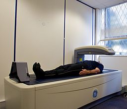
You lie on your back on a couch and are asked to keep still while an X-ray detector (the 'scanner') comes over the area to be tested. An X-ray machine fires X-rays towards the detector. The bones commonly scanned are the bones of the back (the vertebrae), hip and wrist.
Smaller peripheral scanners are available in some places and can be used to check the bone mass density of the heel, wrist or finger.
How long does a DEXA scan take?
The scan usually takes between 5 and 20 minutes, depending on which part of your body is being examined and whether a central or peripheral scanner is being used. There is no 'tunnel' to pass through as there is in other types of scans such as an MRI or CT scan, so it should not affect people who do not like being in enclosed spaces.
DXA scanner
Are DEXA scans safe?
DEXA scans use a very low level of X-ray radiation. This means it is safe for the technician doing the scan to stay in the room with you. (In standard X-ray tests, the technician has to stay behind a protective screen.)
Preparing for a DEXA scan
You do not need to do any special preparation prior to a DEXA scan. You can normally remain fully clothed, although you will need to avoid or remove clothes with metal in them (for example, zips, belts, buttons). You may also be asked to remove jewellery for the scan. In some places, you may be given a gown to wear.
Who should have a DEXA scan?
A DEXA scan may be advised if you have had a fracture of a bone after a minor injury. It may also be advised if you are considered at increased risk of 'thinning' of the bones (osteoporosis) and therefore at increased risk of having a fracture in future.
If your doctor thinks you have risk factors for osteoporosis, they may use b risk calculator such as one called FRAX® or QFracture®. This gives an idea of how likely you are to fracture your bones after a minor knock. If your risk is at a medium level, your doctor would then arrange a DEXA scan. This enables them to gain a clearer picture of your risk and then to decide whether you need any treatment.
DEXA scans are also used to monitor whether treatment for osteoporosis is working.
DEXA scans are not advised for women who are pregnant. You should also not have a DEXA scan within two weeks of certain other types of scans - for example, those using contrast dye.
For further information, see the separate leaflet called Osteoporos.
Patientval för Bilddiagnostik

Tester och undersökningar
Benscanning
En skelettscintigrafi är en speciell typ av nukleärmedicinsk procedur som använder radionuklider för att skapa en bild av skelettet. Radionuklider är kemikalier som avger radioaktivitet som kan upptäckas av speciella skannrar. En skelettscintigrafi skiljer sig från en bentäthetsmätning (DEXA). Se broschyren om DEXA-skanning för mer information om detta bentäthetstest. En DEXA-skanning är också känd som en DXA-skanning. Obs: informationen nedan är endast en allmän vägledning. Arrangemangen och sättet tester utförs på kan variera mellan olika sjukhus. Följ alltid instruktionerna från din läkare eller lokala sjukhus.
av Dr Doug McKechnie, MRCGP

Tester och undersökningar
Bariumlavemang
En bariumröntgen är ett test som används för att hjälpa till att se konturerna av tjocktarmen (kolon). Observera: informationen nedan är endast en allmän vägledning. Arrangemangen och sättet som tester utförs på kan variera mellan olika sjukhus. Följ alltid instruktionerna från din läkare eller ditt lokala sjukhus.
av Dr Colin Tidy, MRCGP
Vanliga frågor
What is the purpose of a DEXA scan?
A DEXA scan is used to measure the density of bones. This measurement helps determine how strong your bones are and how likely they are to break (fracture). It can be advised if you've already had a fracture from a minor injury, or if you're considered at high risk of osteoporosis (thinning of the bones).
Does a bone density test show cancer?
The article states that a DEXA scan measures bone density to assess the risk of fractures or monitor osteoporosis treatment. It does not mention that a DEXA scan is used to detect cancer.
Do I need to do anything special to prepare for a DEXA scan?
You typically don't need any special preparation. You can usually remain fully clothed, but you will need to avoid or remove any clothing with metal, such as zips, belts, or buttons. You might also be asked to take off jewellery, and in some cases, you may be given a gown to wear.
Who should not have a DEXA scan?
DEXA scans are not recommended for pregnant women. Additionally, you should not have a DEXA scan within two weeks of having other types of scans that involve the use of contrast dye.
What is the difference between central and peripheral DEXA devices?
Central DEXA devices are large machines that measure bone density in the core parts of your skeleton, like your hip and spine. Peripheral DEXA devices are smaller, portable machines used for areas like the wrist, heel, or finger. Peripheral devices mainly indicate if further testing is needed, as they are not as accurate as the larger central machines.
Vidare läsning och referenser
- Osteoporos: bedömning av risken för benskörhetsfraktur; NICE Klinisk Riktlinje (augusti 2012, uppdaterad februari 2017)
- Hantering av osteoporos och förebyggande av benskörhetsfrakturer - En nationell klinisk riktlinje; Scottish Intercollegiate Guidelines Network (SIGN - januari 2021)
- Osteoporosis - prevention of fragility fractures; NICE CKS, juli 2021 (endast tillgång i Storbritannien)
- Klinisk riktlinje för förebyggande och behandling av osteoporos; Nationella Osteoporosriktlinjegruppen (uppdaterad september 2021)
Om författarenVisa fullständig biografi
Uppdaterad av

Dr Colin Tidy, MRCGP
Allmänläkare, Medicinsk Författare
MBBS, MRCGP, MRCP (Paediatrics), DCH
Dr Colin Tidy är en NHS-läkare, baserad i Oxfordshire.
Om recensentenVisa fullständig biografi

Dr Hayley Willacy, FRCGP
Allmänläkare, Medicinsk Författare
MBChB (1992), DRCOG, DFFP, MRCOG (Part 1) MRCGP (2007), DFSRH (2013), MSc - medical education (2020)
Dr Hayley Willacy var en NHS-läkare som arbetade i nordvästra England och gick i pension från klinisk praktik 2022 efter 30 år.
Artikelhistorik
Artikeln finns också på Engelska, Tyska, Spanska, Franska, Italienska, Portugisiska, Hindi, Hebreiska, Arabiska, och Svenska.
Nästa granskning förfaller: 18 mars 2028
20 mars 2023 | Senaste versionen
Dela dina tankar eller erfarenheter med andra i vår patientgemenskaper.
Enkel tillgång till pålitliga behandlingar och hälsotjänster online.
Common conditions
Get help with common conditions through the NHS, without a GP appointment.





Anmäl dig till Patientens nyhetsbrev
Din veckovisa dos av tydliga, pålitliga hälsoråd - skrivna för att hjälpa dig känna dig informerad, självsäker och i kontroll.
Genom att prenumerera accepterar du våra Sekretesspolicy. Du kan avsluta prenumerationen när som helst. Vi säljer aldrig dina uppgifter.