Elektroencefalograf
EEG
Granskad av Dr John Cox, MRCGPSenast uppdaterad av Dr Colin Tidy, MRCGPSenast uppdaterad 24 May 2018
Uppfyller patientens redaktionella riktlinjer
- Ladda nerLadda ner
- Dela
- Language
- Diskussion
- Ljudversion
- Lägg till i föredragna källor på Google
I denna serie:EpilepsiTyper av anfallTonisk-kloniska anfallFokala anfallAbsensepilepsiEpilepsimedicin och biverkningar
Innehållsmeddelande
Den här sidan har inte granskats på senare tid, så viss information eller vissa externa länkar kan ha ändrats.
An electroencephalograph is a useful test to help diagnose epilepsy. It records the electrical activity of the brain. However, a normal result does not rule out epilepsy.
Notera: informationen nedan är endast en allmän vägledning. Arrangemangen och sättet tester utförs på kan variera mellan olika sjukhus. Följ alltid instruktionerna från din läkare eller lokala sjukhus.
Överblick
An electroencephalograph (EEG) detects and records the electrical signals of your brain.
It is a painless and harmless test.
Small patches called electrodes are attached to your scalp and connected to a machine.
An EEG may be used to help diagnose conditions like epilepsy.
A normal EEG result does not rule out epilepsy.
An abnormal EEG result does not necessarily mean you have epilepsy.
There are special types of EEG tests, such as sleep EEG and ambulatory EEG, for specific situations.
What is an electroencephalograph?
The brain normally produces tiny electrical signals that come from the brain cells and nerves which send messages to each other. These electrical signals can be detected and recorded by the electroencephalograph (EEG) machine. The EEG test is painless and harmless. (The EEG machine records electrical signals coming från your brain - it does not put any electricity into your brain or body.)
You may be advised to have an EEG if you have had symptoms which may be due to a seizure. (Old words for seizure are convulsion or 'fit'.) An EEG test is useful in diagnosing conditions such as epilepsy. See the separate leaflet called Types of Epilepsy and Seizures.
How is the test done?
When you have the test, the operator will attach several small patches (electrodes) to your scalp. Wires from the electrodes are connected to the EEG machine. The machine detects and amplifies the electrical signals and records them on to a paper or computer. The test takes about 20-30 minutes. The electrodes are removed at the end of the test.
EEG machine
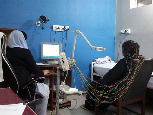
For the duration of the test you may be asked to sit in a chair or lie on a couch. At some point you may be asked to blink lots of times, or to breathe deeply. These can sometimes trigger patterns of electrical activity in the brain which are associated with certain types of epilepsy.
What can the test show?
A normal ('negative') result
This shows a typical pattern of electrical activity from the brain. Most people without epilepsy, and many people with epilepsy, have a normal result. This is because an EEG only shows the electrical activity of the brain when the test is done. With many types of epilepsy, you only have abnormal electrical activity during a seizure. For the rest of the time, the pattern is normal.
An abnormal ('positive') result
This shows abnormal patterns of electrical activity. Some people with certain types of epilepsy have abnormal patterns all the time, not just when they have seizures. (Although, during a seizure the activity is even more abnormal.) For example, children with typical 'absence seizures' often have a characteristic EEG pattern which helps to confirm this type of epilepsy.
However, a small number of people who never have seizures and who don't have epilepsy, have some abnormal patterns of electrical activity in the brain.
Therefore, if you have symptoms which are thought to be seizures, an abnormal EEG means that the diagnosis is likely to be epilepsy. However, a normal result does not rule out epilepsy, and an abnormal result does not necessarily mean that you have epilepsy.
Children and electroencephalography
The interpretation of a child's EEG recording is more difficult. This is because the EEG changes during childhood. An adult pattern is usually developed by the age of 15 years. As the EEG pattern in infants and children can vary considerably, careful interpretation of the test is necessary.
Some specialised types of electroencephalograph test
Strobe lighting
In some cases, a strobe light may be used during an EEG test. This aims to detect if this alters the electrical pattern in the brain. (Usually it does not. However, a small number of people have seizures triggered by flickering or strobe lights and so this may help to identify these people.)
Sleep EEG
In this test, an EEG is performed while you are sleeping. This is usually carried out when you are in hospital. You may need to have one of these if your seizures happen when you are asleep or when you are tired.
Sleep-deprived EEG
There may be a better chance of detecting abnormal brain activity after a period of time when you are deprived of sleep. Therefore, sometimes the EEG test is done after you have stayed awake for all or most of the night. It is done in the same way as the normal test but with you asleep - after the period of 'sleep deprivation'.
Ambulatory EEG
This may be advised in cases where the diagnosis is not clear. It uses a portable EEG machine which records the brain's electrical activity when you are going about your normal activities. The electrodes can usually be hidden under your hair. The wires are connected to a small machine which you wear on a belt (a bit like wearing an mp3 player).
You may be asked to keep a diary when you have meals, go to sleep, and have any symptoms which may be a seizure. The patterns on the EEG can be analysed to see if they change when symptoms occur. It may help to confirm whether certain symptoms are due to seizures.
Video-telemetry
Where there is doubt about a diagnosis of epilepsy, or where the type of seizures someone experiences is unclear, video-telemetry can be helpful. This is a test that uses a video camera linked to an EEG machine.
The camera will visually record your movements and, at the same time, the EEG machine will record your brainwave pattern. Both the video and EEG are stored on to a computer so that they can be reviewed once the test is finished.
The doctor will be able to see any seizures that you may have had, as well as any changes in your EEG at that time. The test is often carried out over a number of days in order to increase the chances of recording one of your seizures.
What should I do to prepare for an electroencephalograph?
Your doctor should give you instructions about what you need to do to prepare for an EEG. Commonly there is no preparation necessary. You should not stop any medication you take for seizures unless advised to by your doctor.
Patientval för Allmänna tester och undersökningar

Tester och undersökningar
Nervledningsstudier
Nerve conduction studies help to test how well and how fast nerves conduct electrical signals. Note: the information below is a general guide only. The arrangements, and the way tests are performed, may vary between different hospitals. Always follow the instructions given by your doctor or local hospital.
av Dr Caroline Wiggins, MRCGP
Tester och undersökningar
Audiologi
Om du eller en familjemedlem misstänker att ni har ett hörselproblem, kommer en audionom vanligtvis att göra en bedömning och kan även vara involverad i att tillhandahålla eventuell nödvändig behandling. Vissa tillstånd som orsakar balanssvårigheter eller yrsel kan uppstå på grund av problem i örat, och audionomer kan utföra specialisttester för att undersöka dessa problem också.
av Dr Surangi Mendis, MRCGP
Vanliga frågor
What is the primary reason for having an EEG test?
An EEG test is primarily used to help diagnose conditions like epilepsy. It's often recommended if you've experienced symptoms that might be seizures, such as convulsions or 'fits'.
How long does an EEG test usually take?
A standard EEG test typically takes about 20-30 minutes to complete. Some specialised types of EEG, like ambulatory EEG or video-telemetry, might last longer or be conducted over several days.
Does having a 'normal' EEG result mean I definitely don't have epilepsy?
Not necessarily. A normal ('negative') EEG result means the electrical activity in your brain appeared typical during the test. For many people with epilepsy, abnormal electrical activity only occurs during a seizure. Therefore, a normal result during an EEG does not rule out epilepsy.
Does an 'abnormal' EEG result always mean I have epilepsy?
No. An abnormal ('positive') EEG result indicates unusual patterns of electrical activity, which can suggest epilepsy, especially if you have symptoms of seizures. However, a small number of people who do not have epilepsy and never experience seizures can also show some abnormal patterns. So, an abnormal result doesn't automatically confirm epilepsy.
Is it safe for children to have an EEG?
Yes, it is safe for children to have an EEG. The test is painless and harmless; it only records electrical signals and does not put electricity into the body. However, interpreting a child's EEG is more complex because brain electrical patterns change significantly during childhood, reaching an adult pattern around 15 years of age.
What is the purpose of flashing a strobe light during an EEG?
In some cases, a strobe light is used during an EEG to see if it triggers changes in the brain's electrical patterns. This is particularly useful for identifying individuals whose seizures are triggered by flickering or strobe lights, though usually, it doesn't alter the electrical pattern.
Vidare läsning och referenser
- Epilepsier: diagnos och behandling; NICE Clinical Guideline (January 2012)
- Diagnos och behandling av epilepsi hos vuxna; Scottish Intercollegiate Guidelines Network – SIGN (2015 – uppdaterad 2018)
- Epilepsi; NICE CKS, mars 2018 (endast tillgång i Storbritannien)
Om författarenVisa fullständig biografi
Uppdaterad av

Dr Colin Tidy, MRCGP
Allmänläkare, Medicinsk Författare
MBBS, MRCGP, MRCP (Paediatrics), DCH
Dr Colin Tidy är en NHS-läkare, baserad i Oxfordshire.
Om recensentenVisa fullständig biografi

Dr John Cox, MRCGP
MA, MB, B Chir, DCH, DRCOG, MRCP (UK), MRCGP
Dr John Cox worked as a Medical Registrar in the UK and New Zealand and as a locum Physician in New Zealand.
Artikelhistorik
Artikeln finns också på Engelska, Tyska, Spanska, Franska, Italienska, Portugisiska, Hindi, Hebreiska, Arabiska, och Svenska.
24 May 2018 | Senaste versionen
Delta i konversationen
Dela dina tankar eller erfarenheter med andra i vår patientgemenskaper.

Fråga, dela, anslut.
Bläddra i diskussioner, ställ frågor och dela erfarenheter inom hundratals hälsorelaterade ämnen.

Känner du dig sjuk?
Bedöm dina symtom online gratis
Anmäl dig till Patientens nyhetsbrev
Din veckovisa dos av tydliga, pålitliga hälsoråd - skrivna för att hjälpa dig känna dig informerad, självsäker och i kontroll.
Genom att prenumerera accepterar du våra Sekretesspolicy. Du kan avsluta prenumerationen när som helst. Vi säljer aldrig dina uppgifter.