Milia
Granskad av Dr Hayley Willacy, FRCGP Senast uppdaterad av Dr Colin Tidy, MRCGPSenast uppdaterad 14 dec 2022
Uppfyller patientens redaktionella riktlinjer
- Ladda nerLadda ner
- Dela
- Language
- Diskussion
- Ljudversion
- Lägg till i föredragna källor på Google
Medicinska yrkesverksamma
Professionella referensartiklar är utformade för att användas av vårdpersonal. De är skrivna av brittiska läkare och baserade på forskningsbevis, brittiska och europeiska riktlinjer. Du kan hitta Milia artikel mer användbar, eller en av våra andra hälsoartiklar.
What are milia?
Milia are very common, benign, keratin-filled epidermoid cysts that occur in individuals of all ages, from infants to elderly persons.1 .
Primary milia arise spontaneously and are typically seen in infants, when they are congenital, but may also occur in children and adults. Primary milia arise on skin bearing vellus hair follicles, most commonly on the face.
Secondary milia result from damage to eccrine ducts.
Milia en plaque and multiple eruptive milia are distinct entities.
How common are milia?1
Milia are common in all ages and both sexes. Congenital milia affect approximately 50% of infants.
Multiple eruptive milia and milia en plaque are extremely rare.
Milia symptoms2
Milia are superficial, uniform, pearly-white to yellowish, domed lesions measuring 1-2 mm in diameter.
Milia most often arise on the face and are particularly prominent on the eyelids and cheeks, but they may occur elsewhere, including the genitalia.
They are usually asymptomatic but may be itchy.
Primary milia
Primary milia in infants occur on the face, especially the cheeks, nose and around the eyes. They may be more widespread on the scalp, face and upper trunk.
Congenital oral inclusion cysts such as those found on the mucosa (Epstein's pearls) and palate (Bohn's nodules) are thought to be the oral counterparts of milia.
Primary milia in children and adults are most often on the eyelids, cheeks, forehead and genitalia. They may clear in a few weeks or persist for months or longer.
Juvenile milia may be present at birth or appear later in life. They may be associated with other genetic disorders.
Milia in a newborn
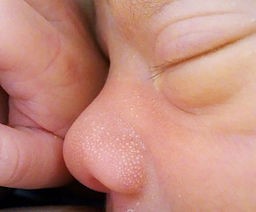
Milia en plaque3
The small papules arise on a distinct, erythematous plaque in the postauricular area, unilaterally or bilaterally.
They are usually seen behind the ears, but may be on the eyelids, cheeks or on the jaw. Submandibular plaques and lesions on the pinna have been reported.4
They especially affect middle-aged women and may be associated with other skin disease - eg, pseudoxanthoma elasticum, discoid lupus erythematosus, lichen planus.2
Multiple eruptive milia
Crops of milia appear over a few weeks to months.5 Most often, they affect the face, upper arms and upper trunk.2
Secondary milia1
These are found anywhere on the body, depending on the predisposing condition:
Disease: post-bullous, typically epidermolysis bullosa and porphyria cutanea tarda but can also occur after other skin conditions - for example, contact dermatitis or lichen sclerosis.
Medication associated with milia includes topical steroids, 5-fluorouracil, ciclosporin and penicillamine.
Traumatic abrasions are a common cause of milia in children. They can also occur following, for example, second-degree burns, dermabrasion or radiotherapy.
Utredningar
The clinical appearance is diagnostic in simple milia and no further investigations are required.
In elderly people with sun-damaged skin, a biopsy may be needed to exclude nodular elastosis of the skin (Favre-Racouchot syndrome). If milia en plaque are suspected, a biopsy is prudent to confirm the diagnosis.
Differentialdiagnos2
Acne vulgaris Closed comedones are more cream than white. They also usually have a small punctum as well as being associated with open (black) comedones.
Syringomas are small papules on lower eyelids and are skin-coloured
Xanthelasma are yellow, flat plaques over the upper or lower eyelids.
Trichoepithelioma.
Follicular (or alopecia) mucinosis.
Milia treatment
Usually no treatment is required.
Some milia may be removed with a needle, as they often shell out easily (this doesn't require anaesthetic).6
Topical peeling agents do not work.
Prognos
They are harmless but can be unsightly. They tend to resolve within a few weeks in infants but may persist in older children and adults.
When to refer milia
For confirmation of diagnosis or reassurance. It is worth referring patients with suspected milia en plaque.
Exklusiva uppdateringar för vårdpersonal
Håll dig informerad med de senaste kliniska uppdateringarna, professionella insikter och evidensbaserad vägledning. Patient Pro-nyhetsbrevet sammanställer viktigt innehåll för vårdpersonal—levererat direkt till din inkorg.
Genom att prenumerera accepterar du våra Sekretesspolicy. Du kan avsluta prenumerationen när som helst. Vi säljer aldrig dina uppgifter.
Vidare läsning och referenser
- O'Connor NR, McLaughlin MR, Ham P; Newborn skin: Part I. Common rashes. Am Fam Physician. 2008 Jan 1;77(1):47-52.
- Berk DR, Bayliss SJ; Milia: a review and classification. J Am Acad Dermatol. 2008 Dec;59(6):1050-63. Epub 2008 Sep 25.
- Milium, milia; DermNet NZ
- Martins LE, Werner B, Fonseca GP; Milia en plaque. An Bras Dermatol. 2010 Nov-Dec;85(6):895-8.
- Hallaji Z, Akhyani M, Jamshidi S, et al; Bilateral retro-auricular milia en plaque: a case report and review of the literature. Dermatol Online J. 2010 Jan 15;16(1):12.
- Batra P, Tsou HC, Warycha M, et al; Multiple eruptive milia. Dermatol Online J. 2009 Aug 15;15(8):20.
- Thami GP, Kaur S, Kanwar AJ; Surgical Pearl: Enucleation of milia with a disposable hypodermic needle. J Am Acad Dermatol. 2002 Oct;47(4):602-3.
Om författarenVisa fullständig biografi
Uppdaterad av

Dr Colin Tidy, MRCGP
Allmänläkare, Medicinsk Författare
MBBS, MRCGP, MRCP (Paediatrics), DCH
Dr Colin Tidy är en NHS-läkare, baserad i Oxfordshire.
Om recensentenVisa fullständig biografi

Dr Hayley Willacy, FRCGP
Allmänläkare, Medicinsk Författare
MBChB (1992), DRCOG, DFFP, MRCOG (Part 1) MRCGP (2007), DFSRH (2013), MSc - medical education (2020)
Dr Hayley Willacy var en NHS-läkare som arbetade i nordvästra England och gick i pension från klinisk praktik 2022 efter 30 år.
Artikelhistorik
Artikeln finns också på Engelska, Tyska, Spanska, Franska, Italienska, Portugisiska, Hindi, Hebreiska, Arabiska, och Svenska.
Nästa granskning: 13 dec 2027
14 dec 2022 | Senaste versionen
Enkel tillgång till pålitliga behandlingar och hälsotjänster online.
Common conditions
Get help with common conditions through the NHS, without a GP appointment.





Mer inom dermatologi
- Furunklar och karbunklar
- Cellulit och rosfeber
- Kontakt- och yrkesdermatit
- Dermatitis artefacta
- Dermatitis herpetiformis
- Dermatofibrom
- Dermatologiska beskrivande termer
- Dermatofytos
- Diabetesfot
- Svampinfektioner i naglarna
- Droppformad psoriasis
- Intradermala och sammansatta nevi
- Perioral dermatit
- Pityriasis versicolor
- Postinflammatorisk hypopigmentering av huden
- Psoriasisartrit
- Schambergs sjukdom
- Skivepitelcancer i huden
- Verrucae
- Gul nagelsyndrom