Mediastinoskopi
Granskad av Dr Colin Tidy, MRCGPSenast uppdaterad av Dr Laurence KnottSenast uppdaterad 17 Feb 2019
Uppfyller patientens redaktionella riktlinjer
- Ladda nerLadda ner
- Dela
- Language
- Diskussion
- Ask Patient
- Ljudversion
- Lägg till i föredragna källor på Google
Innehållsmeddelande
Den här sidan har inte granskats på senare tid, så viss information eller vissa externa länkar kan ha ändrats.
Mediastinoscopy is a type of 'keyhole' surgery that allows doctors to look inside the chest. It can be used to take samples of body tissue for further testing.
Notera: informationen nedan är endast en allmän vägledning. Arrangemangen och sättet tester utförs på kan variera mellan olika sjukhus. Följ alltid instruktionerna från din läkare eller lokala sjukhus.
Överblick
Mediastinoscopy is a 'keyhole' surgery to examine the mediastinum, an area in your chest.
It is often used to diagnose and stage lung cancer and other conditions affecting the chest.
A surgeon inserts a thin tube with a camera through small cuts to see inside your chest.
Small tissue samples can be taken for further examination under a microscope.
You will usually have a general anaesthetic for this procedure and may have a sore throat afterwards.
Contact your doctor if you experience bleeding, fever, severe chest pain, or worsening hoarseness.
What is mediastinoscopy?
Mediastinoscopy involves a small operation to look at an area of the body, called the mediastinum. The mediastinum is part of the chest and contains the heart, gullet (oesophagus), windpipe (trachea) and various nerves. It is the area shown in purple in the image below.
Mediastinum
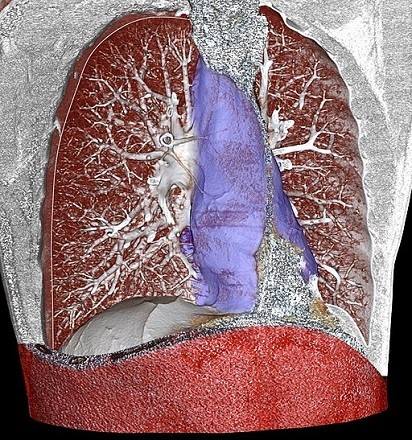
The mediastinum also contains lymph nodes and the thymus. Both of these are part of the immune system and help in fighting infection.
Mediastinoscopy is a type of 'keyhole' surgery where only very small cuts (incisions) are made to the body. It uses a special instrument called a mediastinoscope. This is a thin, tube-like instrument which has a camera built into the end. The 'scope' is inserted into a small incision in the upper chest. This is then linked up to a monitor, allowing the surgeon to see pictures of the chest.
While looking at these pictures, the surgeon may see something which they feel needs investigating further. Using the mediastinoscope they can take very small samples from within the chest. These can then be looked at further under a microscope.
What is mediastinoscopy used for?
The most common reason for mediastinoscopy is as a staging procedure in the diagnosis of non-small cell lung carcinoma. The results obtained from the procedure are very important in tailoring care for the patient.
Other indications for mediastinoscopy include:
The diagnosis and removal of mediastinal masses and enlarged lymph nodes.
Lymphoma, both Hodgkins and non-Hodgkins, can be diagnosed via mediastinoscopy along with sarkoidos which has similar appearances on imaging.
A mediastinal mass, such as a thymoma, may be removed using mediastinoscopy, depending on its size.
A sampling of lymph nodes may diagnose infectious processes such as tuberkulos och fungal infections.
Benign (non-cancerous) mediastinal cysts can be taken out using mediastinoscopy.
Mediastinoscopy is used in the diagnosis and treatment of mesothelioma.
How does mediastinoscopy work?
Before the operation, a small needle will be placed into a vein (usually on the back of the hand) to give you fluids and medication.
Mediastinoscopy is generally done under general anaesthetic, meaning that you will be asleep for the procedure. After you are asleep, a tube (an endotracheal tube - also called an ET tube) will be placed in your throat to help you breathe during the procedure. Your neck and chest will be washed with an antiseptic soap and covered with a sterile drape.
The surgeon may make two or three small cuts (incisions) in the chest wall above the breastbone (sternum). These holes are known as 'ports' and are usually about an inch long. The mediastinoscope is inserted through one hole. The camera in the mediastinoscope feeds video images to a computer screen, allowing the surgeon to see inside the chest.
Your doctor will also insert special surgical instruments through the other ports. He or she will examine the space in your chest between your lungs and heart. Lymph nodes or abnormal tissue samples will be collected and sent to the laboratory for further tests. After the 'scope' is removed from your chest, all these incisions will be closed with a few stitches and covered with a bandage.The entire procedure usually takes about an hour.
What should I do to prepare for mediastinoscopy?
Your doctor should discuss any specific preparation necessary with you before the procedure. Usually there is not much preparation necessary.
Notera: if you take 'blood-thinning' medicines such as warfarin or aspirin, your doctor may ask you to stop taking these for a few days before your operation.
You will also be asked not to eat or drink for a certain length of time before the procedure. Your local hospital will give you information on this.
What can I expect after mediastinoscopy?
Some people may go home after the procedure if the general anaesthesia wears off and they are able to swallow fluids without gagging or choking. Other people may need to stay in hospital for one or two days. If your stitches are not the dissolving type, you will need to return to your doctor to have them removed.
You may have a slightly sore throat - this is normal and usually due to the tube used to help you breathe during the operation. The sites where the cuts (incisions) were made may be sore for a few days after the procedure.
Are there complications after mediastinoscopy?
Most people who have a mediastinoscopy do not develop any complications.
Sometimes the mediastinoscope can temporarily injure a nerve, which may cause hoarseness. This can occasionally be permanent.
In rare cases, bleeding complications might require a transfusion or larger chest surgery. Air leaks from the lung can also occur and occasionally require additional treatment. Usually this involves putting in a drainage tube between your ribs for a few days.
Rarely mediastinoscopy may cause a collapsed lung (pneumothorax) or damage to the gullet (oesophagus).
You should contact your doctor if you become unwell after a mediastinoscopy. In particular, if you have any of the following problems:
Bleeding from your stitches.
Hög temperatur (feber).
Severe chest pain.
Swelling in the neck.
Andnöd.
Difficulty swallowing.
Hoarseness of your voice that lasts more than a few days or continues to worsen.
Advances in X-ray technology and surgical techniques mean that it may be possible to offer you options which cause less discomfort than mediastinoscopy.
Patientval för Bilddiagnostik

Tester och undersökningar
PET-skanning
A PET scan create images which show where cells are particularly active in the body. It is most commonly used to diagnose and assess cancer. Note: the information below is a general guide only. The arrangements, and the way tests are performed, may vary between different hospitals. Always follow the instructions given by your doctor or local hospital.
av Dr Rosalyn Adleman, MRCGP

Tester och undersökningar
CT-skanning
Observera: informationen nedan är endast en allmän vägledning. Arrangemangen och sättet tester utförs på kan variera mellan olika sjukhus. Följ alltid instruktionerna från din läkare eller ditt lokala sjukhus.
av Dr Rachel Hudson, MRCGP
Vanliga frågor
What is the mediastinum, and what organs are located in this area?
The mediastinum is a specific area within the chest that contains several vital organs. These include the heart, the gullet (oesophagus), the windpipe (trachea), and various nerves. It also houses lymph nodes and the thymus, both of which are crucial parts of the immune system, helping the body fight infections.
What is the purpose of the 'ports' in a mediastinoscopy procedure?
During a mediastinoscopy, the surgeon makes two or three small incisions, known as 'ports', in the chest wall just above the breastbone. These ports are typically about an inch long. The mediastinoscope, which has a camera, is inserted through one of these holes. Special surgical instruments are then inserted through the other ports, allowing the surgeon to visualise the inside of the chest and collect tissue samples.
How long does a mediastinoscopy procedure typically last?
The entire mediastinoscopy procedure usually takes approximately one hour to complete.
Are there any specific instructions regarding food and drink before the mediastinoscopy?
Yes, you will be asked not to eat or drink for a certain period before the procedure. Your local hospital will provide you with detailed information about these fasting instructions.
What are some of the signs that I should contact my doctor after a mediastinoscopy?
You should contact your doctor if you experience any of the following problems after a mediastinoscopy: bleeding from your stitches, a high temperature (fever), severe chest pain, swelling in the neck, shortness of breath, difficulty swallowing, or hoarseness that lasts more than a few days or worsens.
Vidare läsning och referenser
- McNally PA, Arthur ME; Mediastinoscopy. StatPearls Publishing. 2018.
- Onat S, Ates G, Avci A, et al; The role of mediastinoscopy in the diagnosis of non-lung cancer diseases. Ther Clin Risk Manag. 2017 Jul 27;13:939-943. doi: 10.2147/TCRM.S144393. eCollection 2017.
Om författarenVisa fullständig biografi
Uppdaterad av

Dr Laurence Knott
Allmänläkare, Medicinsk Författare
BSc (Hons) Biokemi, Läkarexamen
Dr Laurence Knott kvalificerade sig 1973 och har haft omfattande erfarenhet som allmänläkare.
Om recensentenVisa fullständig biografi

Dr Colin Tidy, MRCGP
Allmänläkare, Medicinsk Författare
MBBS, MRCGP, MRCP (Paediatrics), DCH
Dr Colin Tidy är en NHS-läkare, baserad i Oxfordshire.
Artikelhistorik
Artikeln finns också på Engelska, Tyska, Spanska, Franska, Italienska, Portugisiska, Hindi, Hebreiska, Arabiska, och Svenska.
17 Feb 2019 | Senaste versionen
Dela dina tankar eller erfarenheter med andra i vår patientgemenskaper.
Enkel tillgång till pålitliga behandlingar och hälsotjänster online.
Common conditions
Get help with common conditions through the NHS, without a GP appointment.





Anmäl dig till Patientens nyhetsbrev
Din veckovisa dos av tydliga, pålitliga hälsoråd - skrivna för att hjälpa dig känna dig informerad, självsäker och i kontroll.
Genom att prenumerera accepterar du våra Sekretesspolicy. Du kan avsluta prenumerationen när som helst. Vi säljer aldrig dina uppgifter.