Haemangiomata of skin
Peer reviewed by Dr Colin Tidy, MRCGPLast updated by Dr Rosalyn Adleman, MRCGPLast updated 21 Nov 2022
Meets Patient’s editorial guidelines
- DownloadDownload
- Share
- Language
- Discussion
- Audio Version
- Add to preferred sources on Google
Medical Professionals
Professional Reference articles are designed for health professionals to use. They are written by UK doctors and based on research evidence, UK and European Guidelines. You may find one of our health articles more useful.
What are haemangiomata of the skin?
Haemangiomata appear as red to purple papules or plaques with a normal epithelial surface. Compression leads to partial emptying and the colour becomes less prominent. There are various types:1
Capillary naevus
Also known as a naevus simplex this is a small, flat, red or pink patch. Seen on the neck in up to 40% of infants (stork bite), between the eyebrows (angel's kiss) or on the eyelids.1 Stork bites may not fade but are often covered by hair over time. Facial lesions tend to fade in the first year of life.
Port-wine stain
A port-wine stain is a lesion lined with endothelial cells and containing blood vessels. It does not regress with age. It may be associated with Sturge-Weber syndrome (port-wine stain of the face, angiomas of the leptomeninges and choroid, and late glaucoma) and Klippel-Trénaunay syndrome (local overgrowth of soft tissue and bone in an extremity or more extensive area, port-wine stain, varicose veins, cutaneous angiomata and other variable features).2
Vin rose patch
This is a pale pink lesion appearing as a birthmark due to dilatation of the sub-papillary dermal plexus.
Venous lake
These are purple or dark-blue papules caused by dilatation of venules. They present in sun-exposed areas of the body, particularly the lips, faces and ears of elderly patients. The average age at presentation is 65 years. They are probably more common in men than in women. They are of little clinical significance, except that they can be confused with melanomas and pigmented basal cell carcinomas.3
Capillary haemangioma (strawberry naevus)
This is also known as a strawberry naevus or infantile haemangioma. It tends to regress after the first year of life and normally resolves completely after the age of 4 or 5 years. Persistent lesions or those causing obstruction of vision may require treatment.4 5
Cherry angioma
Also known as Campbell de Morgan spots, they appear on the abdomen and chest and are red, slightly elevated keratoangiomata. They do not fade with pressure.6
Telangiectasias
These are permanent dilatations of groups of capillaries or venules. They may be inherited or associated with atopy, sun damage, connective tissue disease, raised oestrogen levels or venous hypertension.7
Haemangiomata diagnosis
The compression test is useful, or the lesion can be examined with a dermatoscope and the blood-filled cavities observed. Sometimes a haemangioma may be confused with a malignant melanoma, if both are dark in colour and of recent origin. Reflectance confocal microscopy, optical coherence tomography, ultrasonography and multispectral imaging are non-invasive imaging techniques that can be used to support clinical examination and dermatoscopy.8 They can be differentiated by excision biopsy. Campbell de Morgan spots (cherry angiomas) are a type of haemangiomata which remain small and increase in number with age. A strawberry mark/naevus is a proliferating haemangioma that occurs in the first year of life and then regresses thereafter.
Venous-lake angiomas are also usually asymptomatic. Women are more likely to present for cosmetic advice or removal. They are soft and compressible. They often have a smooth surface. They are found most often on lips, face, neck and ears. Actinic skin damage often occurs around venous lakes, as they have a shared aetiology.
Lip venous lake
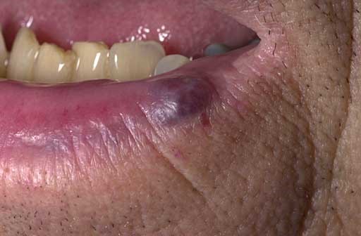
© Dr. Thomas P. Habif M, Creative Commons, via Wikimedia Commons
Haemangiomata primary care treatment
Most haemangiomata require no treatment unless the patient is concerned about their appearance.
Port-wine stains are usually treated by camouflage but the patient may wish to be referred for laser therapy (see 'When to refer for haemangiomata', below).9
Capillary haemangiomata may regress spontaneously. However, if they affect normal development, such as development of binocular vision, or cause bleeding, or obstruction of other organs, or grow rapidly, they may need to be referred for treatment (see 'When to refer for haemangiomata', below).4
Prognosis
This is dependent on the type of haemangiomata. Capillary haemangiomata and capillary naevi usually regress spontaneously, others are more permanent.
When to refer for haemangiomata
Referral should be considered if diagnosis is in doubt or treatment is required. Patients may require laser therapy for port-wine stains. This can be painful but a technique called pneumatic skin flattening, employed during laser treatment has been shown to reduce the discomfort.10
Other treatment options include interferon and surgical excision.11
Exclusive updates for healthcare professionals
Stay informed with the latest clinical updates, professional insights, and evidence-based guidance. The Patient Pro newsletter curates essential content for healthcare professionals—delivered straight to your inbox.
By subscribing you accept our Privacy Policy. You can unsubscribe at any time. We never sell your data.
Further reading and references
- Capillary vascular malformation; DermNet NZ
- Klippel-Trénaunay-Weber image; MedicineNet
- Venous lake; DermNet
- Bang GM, Setabutr P; Periocular capillary hemangiomas: indications and options for treatment. Middle East Afr J Ophthalmol. 2010 Apr;17(2):121-8. doi: 10.4103/0974-9233.63071.
- Infantile haemangioma; DermNet NZ
- Angiomas in adults; DermNet NZ
- Generalised essential telangiectasia; DermNet NZ
- Menge TD, Pellacani G; Advances in noninvasive imaging of melanoma. Semin Cutan Med Surg. 2016 Mar;35(1):18-24. doi: 10.12788/j.sder.2016.003.
- Husain Z, Alster TS; The role of lasers and intense pulsed light technology in dermatology. Clin Cosmet Investig Dermatol. 2016 Feb 4;9:29-40. doi: 10.2147/CCID.S69106. eCollection 2016.
- Kautz G, Kautz I, Segal J, et al; Treatment of resistant port wine stains (PWS) with pulsed dye laser and non-contact vacuum: a pilot study. Lasers Med Sci. 2010 Jul;25(4):525-9. doi: 10.1007/s10103-009-0727-7. Epub 2009 Dec 15.
- Richter GT, Friedman AB; Hemangiomas and vascular malformations: current theory and management. Int J Pediatr. 2012;2012:645678. doi: 10.1155/2012/645678. Epub 2012 May 7.
About the authorView full bio

Dr Rosalyn Adleman, MRCGP
MRCGP
Dr Rosalyn Adleman, is an NHS GP working in north London.
About the reviewerView full bio

Dr Colin Tidy, MRCGP
General Practitioner, Medical Author
MBBS, MRCGP, MRCP (Paediatrics), DCH
Dr Colin Tidy is an NHS Doctor, based in Oxfordshire.
Article history
Article also available in English, German, Spanish, French, Italian, Portuguese, Hindi, Hebrew, Arabic, and Swedish.
Next review due: 20 Nov 2027
21 Nov 2022 | Latest version

Ask, share, connect.
Browse discussions, ask questions, and share experiences across hundreds of health topics.

Feeling unwell?
Assess your symptoms online for free
More in dermatology
- Albinism
- Angio-oedema
- Anogenital warts
- Becker's naevus
- Cellulitis and erysipelas
- Dermatological descriptive terms
- Eczema on hands and feet
- Granuloma annulare
- Halo naevus
- Henoch-Schönlein purpura
- Hidradenitis suppurativa
- Impetigo
- Lichen sclerosus
- Malignant melanoma of skin
- Pemphigus
- Pompholyx
- Shingles
- Smallpox
- Sturge-Weber syndrome
- Xanthelasma