Epidermoid and pilar cysts
Sebaceous cysts
Peer reviewed by Dr Colin Tidy, MRCGPLast updated by Dr Doug McKechnie, MRCGPLast updated 12 Jun 2023
Meets Patient’s editorial guidelines
- DownloadDownload
- Share
- Language
- Discussion
- Audio Version
- Add to preferred sources on Google
Epidermoid and pilar cysts are commonly referred to as 'sebaceous cysts' (pronounced 'seb-ay-shuss'). They are overgrowths of skin cells (called keratin) held together in a little capsule, or sac.
They are harmless smooth lumps just under the surface of the skin. They are not cancerous and do not require removal unless they are bothering you by the look or the feel of them.
They can occur almost anywhere in the body, but are commonly found on the back or scalp.
At a glance
Cysts are sacs in the body filled with fluid or semi-fluid material.
Epidermoid and pilar cysts are common types found under the skin.
Epidermoid cysts often appear on the face, neck, chest, and back.
Pilar cysts commonly develop on the scalp, especially in middle-aged women.
These cysts usually appear as smooth, round, painless lumps under the skin.
Cysts are non-cancerous but can become red, inflamed, and painful if infected.
You should see a doctor if a cyst grows quickly or becomes painful.
What are cysts?
A cyst is a sac that is filled with a fluid or semi-fluid material. Cysts develop in various places in the body and arise from different tissues in the body. Two of the most common types of cyst that occur under the skin surface are epidermoid and pilar cysts.
These cysts are often called sebaceous cysts but this term is no longer correct, as the origin of these cysts is not from the sebaceous glands in the skin, as was once thought.
Many doctors still call them sebaceous cysts, though.
What is an epidermoid cyst?
An epidermoid cyst is a cyst where the cyst sac forms from epidermal cells that normally occur on the top layer of the skin (the epidermis).
There are lots of other names for epidermoid cysts, such as epidermal cysts, inclusion cysts, and keratin cysts.
What is a pilar cyst?
A pilar cyst is a cyst where the cyst sac forms from cells similar to those that are in the bottom of hair follicles (where hairs grow from).
A pilar cyst is also known as a trichilemmal cyst.
In both cases, the semi-fluid content of the cyst looks a bit like cottage cheese. This substance is soggy keratin. Keratin is made by skin cells and is the substance that hairs are made from and the substance that covers the top layer of the skin.
Who gets them?
Epidermoid cysts can affect anyone but are most common in young and middle-aged adults. They can appear anywhere on the skin but develop most commonly on the face, neck, chest, upper back and sometimes on the scrotum.
Pilar cysts can affect anyone but are most common in middle-aged women. They can appear anywhere on the skin but develop most commonly on the scalp. It is common for several to develop at the same time on the scalp.
Epidermoid and pilar cysts symptoms
Epidermoid and pilar cysts are smooth round lumps which you can see and feel just beneath the skin surface. They are very common.
They range in size but often they are small, like a pea. Sometimes, they can grow slowly over many months to become a few centimetres in diameter. They look very similar to each other but can be distinguished from each other if the cells that form the cyst sac are looked at under the microscope.
Epidermoid and pilar cysts usually cause no symptoms. Occasionally:
Cysts can become infected, when they may become red, inflamed and painful. A course of antibiotics will usually clear an infection if it occurs. Sometimes they settle down even without antibiotics.
The cyst may leak the cheese-like material on to the skin if the cyst is punctured or damaged.
A little horn may grow on the skin over the cyst.
A cyst may form in an uncomfortable place such as in the genital skin or beside a nail.
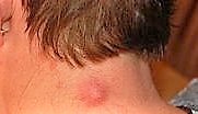
This photo shows the typical appearance of an epidermoid cyst on someone's neck:
Epidermoid cyst on the neck
What causes epidermoid and pilar cysts?
It seems that some cells that are normally near to the surface of the skin (cells of the epidermis or cells in hair follicles) get into deeper parts of the skin and continue to multiply. The cells that multiply form into a sac and produce the keratin that they would normally make on the top layer of the skin. The keratin becomes soggy and forms into a cheese-like substance and a cyst can occur.
Usually epidermoid cysts pop up for no particular reason: there is nothing you can do to prevent them. They are not related to cleanliness, nor will exfoliating stop them occurring.
Pilar cysts on the scalp can be hereditary and run in families, although this is rare.
Rarely, epidermoid cysts can occur due to genetic disorders. A genetic condition might be present if someone has a lot of epidermoid cysts in unusual locations (like on the fingers and toes).
Are epidermoid and pilar cysts painful?
Not usually. If they do not bother you then it is best just to leave them alone. If one has grown recently it's worth asking your doctor to check that it is a harmless cyst. Sometimes a person with an epidermoid or pilar cyst requests that it be removed. This is usually for one of three reasons:
Cosmetic reasons. For example, the cyst is in an obvious site on the skin and looks unsightly.
They are sometimes easy to catch and traumatise. This typically occurs on the scalp when combing hair.
If the cyst often becomes infected or irritating.
Epidermoid and pilar cysts are non-cancerous (benign). They do not spread to other parts of the body or cause any serious problems.
Removal of epidermoid and pilar cysts
If required, the cyst can usually be easily removed by a simple operation under local anaesthetic. The surrounding skin is numbed by injecting some local anaesthetic. A small cut is made over the cyst. It can then be gradually teased out by the doctor. The wound is then stitched up. A small scar will result. Sometimes, after the removal of a cyst, it gradually regrows in the same site under the scar.
Bear in mind you then exchange a small cyst for a scar: some people would rather have been left with the cyst, once they see the results of the surgery.
Patient picks for Skin rashes
Skin, nail and hair health
Hives
Hives (also called urticaria) are itchy red or white bumps on the skin that look like a nettle rash. Hives are common and are often caused by an allergic reaction, stress, infections, or certain medications. Most hives get better on their own within a few days. This leaflet discusses the causes, types, and treatment for hives.
by Dr Philippa Vincent, MRCGP

Skin, nail and hair health
Lichen planus
Lichen planus mainly causes an itchy skin rash. In some people it also affects other parts of the body such as the mouth, genitals, nails and hair. Most people get better in 6-12 months. Treatment, commonly with a steroid cream or ointment, often eases the itch and may reduce the rash until the condition clears.
by Dr Hayley Willacy, FRCGP
Frequently asked questions
Can sebaceous cysts be itchy?
Epidermoid and pilar cysts, sometimes incorrectly called sebaceous cysts, usually do not cause any symptoms. While they can become inflamed, painful, or leak, the article does not mention itchiness as a typical symptom.
Do pilar cysts keep growing?
Epidermoid and pilar cysts can grow slowly over several months, sometimes reaching a few centimeters in diameter.
How to prevent scalp cysts?
The article states that epidermoid cysts usually appear for no particular reason, and there is nothing specific you can do to prevent them. They are not linked to cleanliness or exfoliating. While pilar cysts on the scalp can sometimes be hereditary, the article does not provide methods for prevention.
Further reading and references
- Cysts; DermNet NZ
- Jawa DS, Sircar K, Somani R, et al; Gorlin-Goltz syndrome. J Oral Maxillofac Pathol. 2009 Jul;13(2):89-92. doi: 10.4103/0973-029X.57677.
- Ali SY, Prabhat S, Ramanamurty ChV, et al; Coexistence of porokeratosis of Mibelli with Gardner's syndrome: A rare case report. Indian Dermatol Online J. 2011 Jul;2(2):94-6. doi: 10.4103/2229-5178.86001.
- Verma S, Kushwaha JK, Sonkar AA, et al; Giant sublingual epidermoid cyst resembling plunging ranula. Natl J Maxillofac Surg. 2012 Jul;3(2):211-3. doi: 10.4103/0975-5950.111386.
About the authorView full bio
Updated by

Dr Doug McKechnie, MRCGP
Medical Writer
MA, MBBS, MSc, DRCOG, MRCP(UK), MRCGP(2021), FHEA
Dr Doug McKechnie is an NHS GP working in London. He works full-time clinically and is also the Deputy Lead for the Clinical and Professional Practice module at University College London Medical School.
About the reviewerView full bio

Dr Colin Tidy, MRCGP
General Practitioner, Medical Author
MBBS, MRCGP, MRCP (Paediatrics), DCH
Dr Colin Tidy is an NHS Doctor, based in Oxfordshire.
Join the conversation
Share your thoughts or experiences with others in our patient communities.

Ask, share, connect.
Browse discussions, ask questions, and share experiences across hundreds of health topics.

Feeling unwell?
Assess your symptoms online for free
Sign up to the Patient newsletter
Your weekly dose of clear, trustworthy health advice - written to help you feel informed, confident and in control.
By subscribing you accept our Privacy Policy. You can unsubscribe at any time. We never sell your data.