X-ray test
Peer reviewed by Dr Colin Tidy, MRCGPLast updated by Dr Hayley Willacy, FRCGP Last updated 21 Aug 2023
Meets Patient’s editorial guidelines
- DownloadDownload
- Share
- Language
- Discussion
- Audio Version
- Add to preferred sources on Google
X-ray tests show bones and certain other tissues.
At a glance
X-rays are high-energy radiation used to create images of the inside of the body.
Dense tissues like bone appear white, while air-filled areas appear black on an X-ray picture.
X-rays can help diagnose issues with bones, joints, the heart, lungs, and gut.
The test is painless, but you must stay still for a clear picture.
Repeated X-rays carry a small risk of cell damage.
Pregnant women should avoid X-rays due to a small risk to the baby.
A radiologist interprets the X-ray, and a report is sent to your doctor.
What are X-rays?
X-rays are a type of high-energy radiation. An X-ray machine can produce short bursts of X-rays. The rays pass easily through fluids and soft tissues of the body. However, dense tissue such as bone will block some of the X-rays. (Density means how much of something there is in a certain amount of space.) The more dense the tissue, the fewer X-rays pass through. Air and water are less dense because the particles which make them are not held closely together. They let more X-rays pass through them.
What do chest X-rays show?
In an X-ray picture bones show as light/white areas, whereas air shows as black or darker areas. The other lighter area in the middle of the image is the heart. The muscle which makes up the heart is also quite dense and stops X-rays passing through.
Posteroanterior chest X-ray
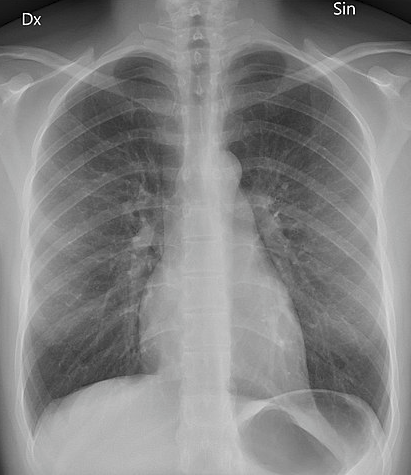
© Mikael Häggström, CC0, via Wikimedia Commons
How is an X-ray test done?
A film, similar to a photographic film, is placed behind the part of the body being X-rayed. The X-ray machine fires a short burst of X-rays through part of your body. The X-rays hit the film, which is then developed. The more X-rays that hit the film, the blacker it develops. So, dense parts of the body that block many of the X-rays show up as white (such as bones). Hollow or air-filled parts of the body show up as black (such as parts of the lung). Soft tissues (such as muscle and body organs) show up as various shades of grey, depending on how dense they are.
The developed film is studied by an X-ray doctor (radiologist) who sends a report to the doctor who requested the test.
An ordinary X-ray test is painless. You cannot see or feel X-rays. You should stay still when the X-ray beam is 'fired', as otherwise the picture may be blurred.
What can an X-ray diagnose?
Bones
Bones, teeth, bone fractures and other abnormalities of bone.
Joint spaces and some abnormalities of joints, such as osteoarthritis.
Chest
The size and shape of the heart. So, certain heart conditions can be detected.
Changes in the density of some softer tissues. For example, a lung tumour is more dense than air-filled lung and will show as a 'shadow' on a chest X-ray. A breast tumour (breast cancer) is more dense than ordinary breast tissue and shows as a 'shadow' on an X-ray of the breast. An X-ray of the breast is also known as a mammogram.
Lung infections (such as pneumonia or tuberculosis) can be diagnosed with a chest x-ray.
Collections of fluid - for example, in the lung or gut - may show as grey 'shadows' against the normal black of the air-filled chest, or hollow gut.
Brain
Blocked blood vessels - a contrast agent that contains iodine can help make areas of your circulatory system (blood vessels) visible on X-rays. A blocked blood vessel in the brain occurs in a stroke. This type of x-ray is called an angiogram.
Abdomen
Digestive tract problems (such as difficulty swallowing), are investigated by a barium swallow. A drink containing a contrast agent called barium, is swallowed and this shows up the outline of the gullet. Most blockages can be highlighted this way.
Accidentally swallowed items - metal, glass and stone objects show up quite well on plain x-rays. They will show the doctors where the object is and allow planning of how best to remove it. Other items (such as those made of wood, or plastic) may be seen best using other methods of imaging such as ultrasound or CT scanning.
Types of X-rays
An ordinary (often called 'plain') X-ray is a quick, easy and relatively cheap test. It may be all that is needed to diagnose or assess various problems. However, an ordinary X-ray has limited uses.
More specialised types of x-rays include:
Contrast studies: substances are used to highlight certain structures in the x-rays. Common examples are blood vessels (angiograms), different parts of the gut (barium swallows and enemas), and urinary system including the bladder and kidneys, (urograms).
Mammograms: these are special x-rays of the breasts designed to show possible cancerous areas. They are used in breast screening programmes.
CT (computerised tomography); a series of x-ray pictures from different angles which a computer puts togther to give a detailed whole picture.
DEXA (dual energy x-rays absorptiometry); a low dose of x-rays which is designed to measure bone density. This is useful when diagnosing osteoporosis.
How do I get an X-ray?
Your GP or consultant may order an X-ray to help diagnose a medical problem you have been discussing with them. X-rays are usually taken in your local hospital department, although some larger health centres also now take certain simple X-rays.
A report on the X-ray is sent to the doctor who ordered the test. The report will come from a specialist in X-rays and their interpretation - a radiologist. The pictures obtained from the X-ray are not usually seen by the requesting doctor, unless it is an emergency situation such as in an A&E department.
How long do X-ray results take?
X-ray pictures are looked at by a specialist called a radiologist, who sends a report to the requesting doctor. This should be available to that doctor within 1-2 weeks.
If there is a serious or urgent finding the report is sent as soon as possible.
Are X-rays dangerous?
There is very little risk with having one X-ray test. However, with repeated tests there is a risk that the X-rays may damage some cells in the body, possibly leading to cancer in the future. The amount of radiation is always kept to the minimum needed to obtain a good picture of the particular body part being checked. (Also, radiographers who take the X-ray pictures always wear lead aprons or go behind a protective screen when the X-rays are fired, to avoid repeated radiation exposure to X-rays.)
Can you have an X-ray while pregnant?
If possible, pregnant women should not have an X-ray test, as there is a small risk that X-rays may cause an abnormality to the unborn child. This is why women are asked if they are, or might be, pregnant, before having an X-ray.
Patient picks for Imaging

Tests and investigations
Barium enema
A barium enema is a test used to help see the outline of the large bowel (colon). Note: the information below is a general guide only. The arrangements, and the way tests are performed, may vary between different hospitals. Always follow the instructions given by your doctor or local hospital.
by Dr Colin Tidy, MRCGP

Tests and investigations
CT scan
Note: the information below is a general guide only. The arrangements, and the way tests are performed, may vary between different hospitals. Always follow the instructions given by your doctor or local hospital.
by Dr Rachel Hudson, MRCGP
Frequently asked questions
What is the typical time frame for receiving my X-ray results?
A specialist called a radiologist will examine your X-ray pictures and send a report to your doctor. This report should typically be available to your doctor within one to two weeks.
Why would my doctor order an X-ray instead of another type of scan?
An ordinary or 'plain' X-ray is a quick, easy, and relatively inexpensive test that can often provide enough information to diagnose or assess a variety of problems. While it has limitations, it can be a suitable first step in many situations.
What is the role of a radiologist in the X-ray process?
A radiologist is an X-ray doctor who specialises in studying and interpreting the developed X-ray film. They then send a report with their findings to the doctor who originally requested the test.
How are images from an X-ray different from other types of scans?
In an X-ray, dense tissues like bones appear as light or white areas, while air shows up as black. Soft tissues show up in various shades of grey. This contrast helps identify issues based on density differences within the body.
If I have an X-ray, will I be able to see the pictures myself?
The X-ray pictures are usually not seen by the doctor who requested the test, unless it is an emergency situation, such as in an A&E department. The radiologist analyses them and sends a report to your doctor.
Further reading and references
- Tafti D, Maani CV; X-ray Production.
- Frane N, Bitterman A; Radiation Safety and Protection.
About the authorView full bio

Dr Hayley Willacy, FRCGP
General Practitioner, Medical Author
MBChB (1992), DRCOG, DFFP, MRCOG (Part 1) MRCGP (2007), DFSRH (2013), MSc - medical education (2020)
Dr Hayley Willacy was an NHS GP working in northwest England, who retired from clinical practice in 2022 after 30 years.
About the reviewerView full bio

Dr Colin Tidy, MRCGP
General Practitioner, Medical Author
MBBS, MRCGP, MRCP (Paediatrics), DCH
Dr Colin Tidy is an NHS Doctor, based in Oxfordshire.
Article history
The information on this page is written and peer reviewed by qualified clinicians.
Article also available in English, German, Spanish, French, Italian, Portuguese, Hindi, Hebrew, Arabic, and Swedish.
Next review due: 19 Aug 2028
21 Aug 2023 | Latest version
Join the conversation
Share your thoughts or experiences with others in our patient communities.

Ask, share, connect.
Browse discussions, ask questions, and share experiences across hundreds of health topics.

Feeling unwell?
Assess your symptoms online for free
Sign up to the Patient newsletter
Your weekly dose of clear, trustworthy health advice - written to help you feel informed, confident and in control.
By subscribing you accept our Privacy Policy. You can unsubscribe at any time. We never sell your data.
More in tests and investigations
- Antibody and antigen tests
- Biopsy
- Bowel cancer screening
- Cardiac catheterisation
- Cardiac enzymes
- Electrocardiogram
- Endometrial biopsy
- ERCP
- Home and ambulatory blood pressure recording
- Ketones in urine
- Liver function tests
- Lumbar puncture
- Midstream specimen of urine
- Patch testing for contact dermatitis
- Radionuclide scan
- Routine kidney function blood test
- Sigmoidoscopy
- Synacthen test
- Thyroid function tests
- Ultrasound scan